The role of neuroimaging in diagnosing Alzheimer's disease
Explore the power of imaging techniques in unraveling the mysteries of Alzheimer's, and learn how they are paving the way for more accurate diagnosis.
Alzheimer’s disease, a progressive neurological disorder, manifests insidiously, stealthily eroding memories, cognitive abilities, and, ultimately, identity. While the disease remains incurable, early diagnosis is pivotal in managing symptoms and planning appropriate care. In recent years, medical imaging has emerged as a potent tool for unraveling the mysteries of Alzheimer’s. This article delves into the diverse modalities of medical imaging and their pivotal role in diagnosing Alzheimer’s disease.
Understanding Alzheimer’s Disease
Alzheimer’s disease, the most common form of dementia, typically begins with subtle memory lapses and progresses to severe cognitive impairment. Characterized by the accumulation of abnormal protein deposits, including beta-amyloid plaques and tau tangles, Alzheimer’s inflicts widespread damage on brain cells, disrupting communication among neurons and leading to irreversible cognitive decline.
The Importance of Early Diagnosis for Dementia Using Brain Scans
Early detection of Alzheimer’s disease empowers patients and caregivers to proactively manage symptoms, access appropriate support services, and participate in clinical trials.
Moreover, early intervention strategies, such as lifestyle modifications and medication, can potentially mitigate disease progression and enhance quality of life.
This section explores various diagnostic tools employed in Alzheimer’s diagnosis, ranging from neuropsychological assessments to brain MRI to molecular brain imaging.
Neuropsychological Assessments:
Neuropsychological assessments evaluate cognitive functions such as memory, attention, language, and executive abilities. These tests, administered by trained clinicians, provide valuable baseline data and identify subtle cognitive changes indicative of Alzheimer’s disease.
Standardized instruments such as the Mini-Mental State Examination (MMSE) and Montreal Cognitive Assessment (MoCA) are commonly used to assess cognitive impairment and track disease progression.
Magnetic Resonance Imaging (MRI):
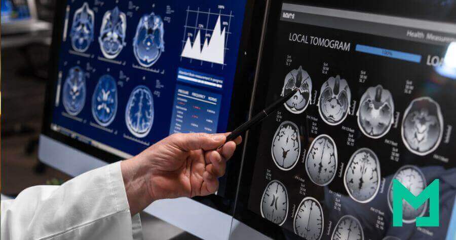
MRI is a non-invasive imaging technique that generates detailed, high-resolution images of the brain’s structure. In Alzheimer’s disease, MRI reveals characteristic patterns of cortical atrophy, particularly in regions associated with memory and cognition, such as the hippocampus and entorhinal cortex. These structural changes serve as important biomarkers for disease progression and severity.
Positron Emission Tomography (PET):
PET imaging allows clinicians to visualize metabolic activity and molecular changes within the brain. In Alzheimer’s diagnosis, PET scans utilizing radiotracers, such as fluorodeoxyglucose (FDG) and amyloid-binding ligands, offer valuable insights. FDG-PET highlights areas of reduced glucose metabolism, indicating neuronal dysfunction and synaptic loss, while amyloid PET imaging detects the accumulation of beta-amyloid plaques, a hallmark feature of Alzheimer’s pathology.
SPECT imaging utilizes radioactive tracers to assess cerebral blood flow and neuroreceptor activity. In Alzheimer’s diagnosis, SPECT scans provide complementary information to PET imaging, revealing regional hypoperfusion and neurotransmitter abnormalities. By mapping perfusion deficits, SPECT helps differentiate Alzheimer’s disease from other types of dementia and contributes to an accurate diagnosis.
Functional MRI (fMRI):
fMRI measures changes in blood oxygenation levels to map brain activity during cognitive tasks. In Alzheimer’s research, fMRI studies elucidate functional connectivity networks and identify aberrant neural circuits associated with disease progression. By probing cognitive domains such as memory, attention, and language, fMRI enhances our understanding of the neurobiological substrates underlying Alzheimer’s pathology.
Diffusion Tensor Imaging (DTI):
DTI is a specialized MRI technique that maps the diffusion of water molecules along white matter tracts in the brain. In Alzheimer’s disease, DTI reveals alterations in white matter integrity, reflecting neuronal degeneration and disconnection. By quantifying changes in diffusion parameters, DTI provides valuable insights into microstructural changes associated with disease pathology.
Blood Tests:
Blood tests offer a minimally invasive approach to assessing biomarkers associated with Alzheimer’s pathology. While no blood test can definitively diagnose Alzheimer’s disease, emerging blood-based biomarkers, including amyloid-beta and tau proteins, show promise for detecting early-stage disease and monitoring progression. Blood tests also evaluate other potential contributors to cognitive decline, such as vitamin deficiencies, thyroid dysfunction, and inflammatory markers.
Spinal Fluid Tests:
A lumbar puncture, or spinal tap, enables the collection of cerebrospinal fluid (CSF) for biochemical analysis. CSF biomarkers, including amyloid-beta, tau, and phosphorylated tau proteins, reflect underlying Alzheimer’s pathology and neuronal injury. Spinal fluid tests provide valuable diagnostic information, particularly in research and specialized clinical settings, aiding in the differential diagnosis of Alzheimer’s disease and other forms of dementia.
Molecular Brain Imaging:
Emerging molecular imaging techniques target specific pathological proteins implicated in Alzheimer’s disease, including beta-amyloid and tau aggregates. PET imaging with amyloid and tau tracers allows visualization of protein deposition in the living brain, facilitating early detection and monitoring of Alzheimer’s pathology.
Molecular brain imaging holds promise for personalized medicine approaches and the development of targeted therapies aimed at modulating disease progression.
Challenges and Limitations
Despite its promise, medical imaging in Alzheimer’s disease faces several challenges and limitations. Variability in imaging protocols, interpretation criteria, and inter-rater reliability may impact diagnostic accuracy and reproducibility. Moreover, the high cost and accessibility constraints associated with advanced imaging modalities pose barriers to widespread adoption, particularly in resource-limited settings.
Future Directions
Advances in medical imaging technology hold promise for refining Alzheimer’s diagnosis and monitoring disease progression. Novel imaging biomarkers, including functional connectivity metrics, synaptic density imaging, and tau PET tracers, are under active investigation to improve diagnostic accuracy and prognostic value. Additionally, machine learning algorithms and artificial intelligence-driven approaches are being developed to analyze complex imaging data in brain scans and identify early signatures of Alzheimer’s pathology with unprecedented precision.
Medical imaging has revolutionized our understanding of Alzheimer’s disease, offering a window into the intricate changes occurring within the brain.
From structural alterations to functional disruptions and molecular signatures, diverse imaging modalities provide invaluable insights for early diagnosis, disease staging, and therapeutic monitoring.
As research continues to unravel the complexities of Alzheimer’s pathology, medical imaging remains at the forefront, guiding clinicians and researchers on the quest to conquer this formidable neurological disorder
Frequently Asked Questions
Is MRI or CT scan better for dementia?
When diagnosing dementia, both MRI (Magnetic Resonance Imaging) and CT (Computed Tomography) scans play crucial roles, but they serve different purposes. MRI scans provide detailed images of the brain’s structure, allowing doctors to detect changes associated with dementia, such as shrinkage of brain tissue or the presence of lesions. On the other hand, CT scans offer a quicker evaluation of brain structure and can help identify large abnormalities like tumors or bleeding.
In the context of dementia diagnosis, MRI scans are generally preferred over CT scans due to their higher sensitivity in detecting subtle changes in brain structure associated with conditions like Alzheimer’s disease. Additionally, MRI can better visualize brain regions involved in memory and cognitive function, often affected in dementia.
However, CT scans may still be used in certain situations, such as when MRI is contraindicated or when a rapid assessment of brain structure is needed. Ultimately, the choice between MRI and CT scans depends on the specific clinical scenario and the patient’s needs. In many cases, a combination of imaging tests, along with mental ability tests, neurological exams, and other diagnostic evaluations, is utilized to diagnose dementia and its underlying causes comprehensively.
What does dementia look like on a CT scan?
Dementia typically does not have specific visual manifestations on a CT (Computed Tomography) scan, as it primarily reveals structural abnormalities in the brain rather than functional changes associated with dementia. However, CT scans may show indirect signs suggesting dementia, such as brain atrophy (shrinkage), enlarged ventricles (fluid-filled spaces in the brain), and white matter changes.
These findings are not exclusive to dementia and can also be seen in other conditions affecting the brain, such as Alzheimer’s disease or vascular dementia. Additionally, CT scans are less sensitive than MRI (Magnetic Resonance Imaging) scans in detecting subtle changes in brain structure associated with dementia.
To diagnose dementia, doctors typically rely on clinical assessments, cognitive tests, and brain imaging tests such as MRI or PET (Positron Emission Tomography) scans, which can provide more detailed information about brain function and detect specific dementia-associated biomarkers.
In summary, while CT scans may reveal general structural changes in the brain that could suggest the presence of dementia, they are not the primary imaging modality used for diagnosing or evaluating the condition.
What is the best test to detect dementia?
The best test to detect dementia typically involves a combination of approaches tailored to the patient’s symptoms, medical history, and clinical presentation. While there is no single definitive test for dementia, a comprehensive diagnostic evaluation may include:
Clinical assessments: Doctors evaluate symptoms reported by the patient and observe their cognitive and behavioral functions during a physical examination.
Neuropsychological tests: These tests assess various cognitive functions such as memory, language, attention, and problem-solving abilities, providing insights into the extent and nature of cognitive impairment.
Brain imaging tests: MRI (Magnetic Resonance Imaging) and PET (Positron Emission Tomography) scans can reveal structural and functional changes in the brain associated with dementia. MRI scans offer detailed images of brain structures, while PET scans can detect abnormalities in brain metabolism or the presence of beta-amyloid plaques, a hallmark of Alzheimer’s disease.
Laboratory tests: Blood tests may be conducted to rule out other potential causes of cognitive impairment, such as vitamin deficiencies, thyroid dysfunction, or infections.
Clinical history and caregiver input: Gathering information about the patient’s medical history, medication use, and changes in behavior or cognition from caregivers or family members can provide valuable insights into the progression of symptoms.
Isaac Health is Medicai’s trusted partner specializing in both early and late-onset Alzheimer’s disease. Schedule an appointment today to access expert assessment and personalized care: https://www.myisaachealth.com/
Experienced marketer, she is interested in health equity, patient experience and value-based care pathways. She believes in interoperability and collaboration for a more connected healthcare industry.