How Neuroimaging changes diagnosis of neurological disorders
Medical imaging is a crucial tool in the field of neurology, as it allows for the visualization of the structures and functions of the brain and nervous system.
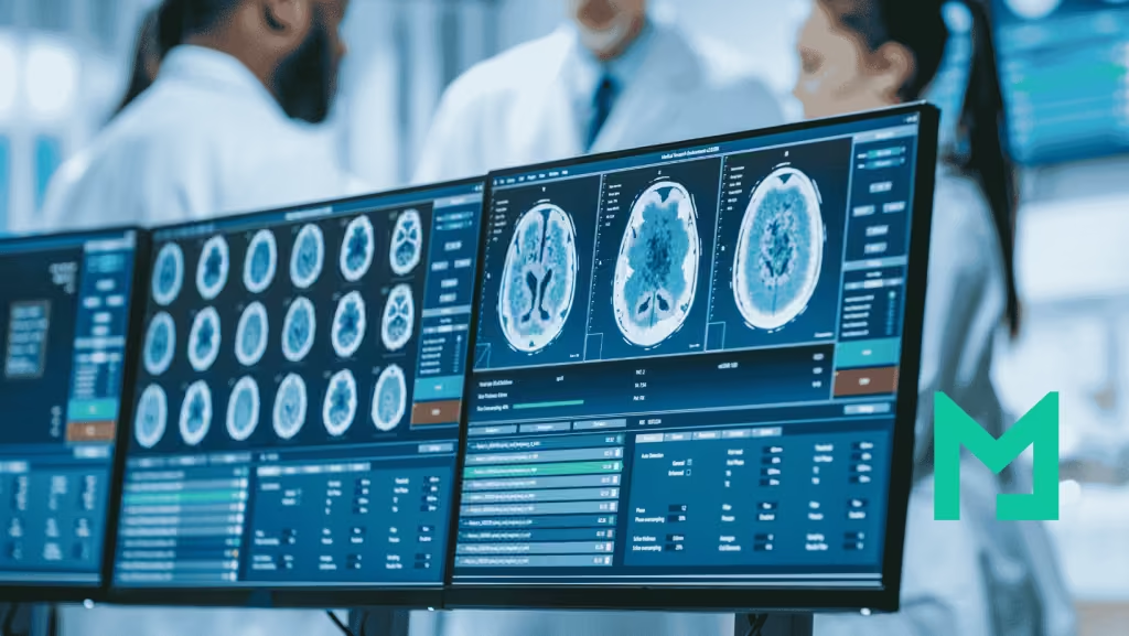
Medical imaging is a crucial tool in the field of neurology, as it allows for the visualization of the structures and functions of the brain and nervous system. These images can help clinicians diagnose and treat a wide range of neurological disorders, including stroke, brain tumors, and multiple sclerosis, among others. In this blog post, we will explore the importance of medical imaging in neurology and how it has revolutionized the field.
Commonly used neuro-imaging techniques
Magnetic Resonance Imaging (MRI)
Neuroimaging techniques have evolved significantly over the past few decades, with new technologies emerging that provide increasingly detailed and accurate images of the brain and its functions. One of the most commonly used neuroimaging techniques is magnetic resonance imaging (MRI), which uses a strong magnetic field and radio waves to produce detailed images of the brain and nervous system. MRI is particularly useful in diagnosing and monitoring conditions such as brain tumors, stroke, and Alzheimer’s disease, among others.
Computed Tomography (CT)
Another widely used neuroimaging technique is computed tomography (CT) scanning, which uses X-rays to create detailed images of the brain and other parts of the body. CT scans are particularly useful in diagnosing acute conditions such as bleeding in the brain, and can also be used to monitor the progression of conditions such as brain tumors.
Positron Emission Tomography (PET)
Positron emission tomography (PET) scanning is another valuable tool in neurology, as it allows clinicians to visualize the metabolic activity of the brain. PET scanning is particularly useful in diagnosing and monitoring conditions such as Alzheimer’s disease and Parkinson’s disease, among others.
Functional Magnetic Resonance Imaging (fMRI)
Functional magnetic resonance imaging (fMRI) is another important neuroimaging technique that allows clinicians to visualize changes in blood flow to the brain in response to different stimuli. This can be particularly useful in diagnosing and monitoring conditions such as epilepsy and multiple sclerosis, among others.
Diffusion Tensor Imaging (DTI)
In addition to these commonly used neuroimaging techniques, there are a number of emerging technologies that are revolutionizing the field of neurology. One of these is diffusion tensor imaging (DTI), which allows for the visualization of the white matter tracts that connect different regions of the brain. DTI is particularly useful in diagnosing and monitoring conditions such as traumatic brain injury and stroke.
Magnetoencephalography (MEG)
Another emerging technology is magnetoencephalography (MEG), which allows for the visualization of the magnetic fields generated by the electrical activity of the brain. MEG is particularly useful in diagnosing and monitoring conditions such as epilepsy and brain tumors, among others.
Key benefits of medical imaging in neurology
The importance of medical imaging in neurology cannot be overstated, as it allows clinicians to visualize the structures and functions of the brain and nervous system in unprecedented detail. This has revolutionized the field of neurology, allowing for more accurate diagnoses and more effective treatments for a wide range of neurological disorders.
One of the key benefits of medical imaging in neurology is the ability to diagnose conditions at an earlier stage, before they become more severe and difficult to treat. For example, MRI can be used to detect brain tumors at an early stage, when they are still relatively small and can be treated more effectively.
Another benefit of medical imaging in neurology is the ability to monitor the progression of conditions over time, allowing clinicians to adjust treatments as necessary. For example, fMRI can be used to monitor changes in brain activity in response to treatment for conditions such as depression and anxiety.
In addition to these benefits, medical imaging in neurology can also help to identify new targets for drug development and other treatments. For example, PET scanning can be used to identify changes in the brain associated with Alzheimer’s disease, which can help researchers to develop new treatments that target these changes.
Future of neuro-imaging
The future of neuroimaging is bright, with new technologies and techniques emerging that promise to provide even more detailed and accurate images of the brain and nervous system. These advances are likely to have a significant impact on the diagnosis and treatment of a wide range of neurological conditions.
Artificial Intelligence (AI)
One area of research that is likely to play an increasingly important role in neuroimaging is artificial intelligence (AI). AI algorithms can be trained to analyze large datasets of neuroimaging data and identify patterns that are difficult for human clinicians to detect. This has the potential to improve the accuracy of diagnoses and identify new treatment targets for neurological conditions.
High-resolution fMRI techniques
Another promising area of research is the development of new imaging technologies that provide even more detailed and accurate images of the brain and nervous system. For example, high-resolution fMRI techniques are being developed that allow for the visualization of brain activity at the level of individual neurons. This could provide unprecedented insights into the workings of the brain and lead to new treatments for neurological conditions.
Wearable sensors
In addition to these advances in imaging technology, there is also growing interest in the use of wearable sensors and other devices that can monitor brain activity in real-time. These devices can provide clinicians with a wealth of data on how the brain responds to different stimuli, which could be used to develop more effective treatments for neurological conditions.
Conclusion
One of the key challenges facing the field of neuroimaging is the development of techniques that can be used in a clinical setting. Many of the emerging neuroimaging technologies are still in the early stages of development and may not be widely available for some time. Additionally, there are challenges associated with interpreting and analyzing large amounts of neuroimaging data, which will require new tools and algorithms to be developed.
Despite these challenges, the future of neuroimaging is full of promise. With new technologies and techniques emerging all the time, the field is likely to continue to grow and evolve in the coming years. This is good news for patients and clinicians alike, as it promises to provide new insights into the workings of the brain and new treatments for neurological conditions.
Experienced marketer, she is interested in health equity, patient experience and value-based care pathways. She believes in interoperability and collaboration for a more connected healthcare industry.