How Healthcare Teams Use Imaging to Guide Patient Rehabilitation

The importance of rehabilitation in healthcare cannot be overstated. The World Health Organization (WHO) reports that an estimated 2.6 billion people worldwide experience a health condition that may require rehabilitation.
The demand for rehabilitation is expected to increase due to the rising prevalence of noncommunicable diseases and population aging. With that, a dire need to translate clinical information into appropriate functional goals is also implied.
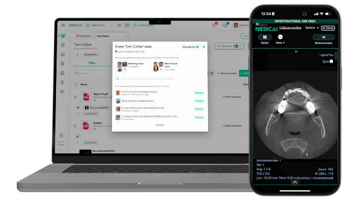
Medical imaging can provide a crucial part of that clinical picture. Structural changes revealed on X-ray, MRI, or CT scans can help healthcare professionals characterize an injury, understand the extent of tissue/bone involvement, and evaluate changes over time. However, effective rehabilitation is only possible when medical imaging findings are placed within the larger context of recovery.
This article will take a closer look at the ways healthcare teams use medical imaging to guide patient rehabilitation.
Understanding the Nature and Extent of the Injury
Clinical examination provides important information about pain, range of motion, swelling, and functional limitations. Medical imaging adds another dimension by allowing clinicians to visualize structural changes that physical examination alone cannot reveal.
So, different imaging modalities answer different clinical questions to determine the condition. For instance, X-rays are especially useful for assessing bones, fractures, and certain joint changes. On the other hand, MRI can provide detailed views of soft tissues such as tendons, ligaments, cartilage, muscles, and bone marrow.
An X-ray may show whether a fracture is displaced, whereas an MRI may reveal damage to a muscle or ligament. Moreover, changes occurring after the initial injury can also be noted. Using imaging, clinicians can identify tissue damage, fluid accumulation, degenerative changes, and signs of healing.
A 2025 study examined the use of handheld musculoskeletal ultrasound by the US Army’s physical therapists during a six-month deployment. The therapists performed 68 tests, and the results led them to modify care plans in 54.4% of cases. The ultrasound information was used to tailor rehabilitation interventions and support return-to-duty decisions.
At the same time, it’s important to interpret imaging results carefully. We can understand this through a 2026 population-based study published in JAMA Internal Medicine. 602 adults aged 41-76 were assessed using bilateral 3-Tesla shoulder MRI and clinical tests.
At least one rotator cuff abnormality was found in 98.7% of participants, including 96% of asymptomatic shoulders and 98% of symptomatic shoulders. These results do not make the shoulder MRI less useful. In fact, they highlight why an image cannot be interpreted independently of the patient.
So, if an MRI shows a rotator cuff abnormality, healthcare providers still need to determine whether it correlates with the patient’s pain, weakness, and limited mobility. Hence, imaging works best as one component of a larger clinical assessment. Its findings can help:
- Identify affected structures and characterize underlying injury
- Recognize precautions that may influence movement
- Understand healing or structural changes during recovery
- Place functional findings in context, so they are not interpreted in isolation
- Establish realistic rehabilitation goals based on the patient’s condition and stage of recovery
Turning Imaging Findings Into a Personalized Rehabilitation Plan
In the previous point, we touched upon recovery and how imaging supported it. It is equally important to translate imaging results into functional implications. A scan primarily shows a fracture, ligament injury, neurological lesion, or other structural change.
Then, the rehabilitation team must determine what that finding means for the patient’s ability to move, perform tasks, and participate in daily life. Different members of the care team must bring in their perspectives to get a clearer picture of the patient. For instance, physicians may use imaging to establish a diagnosis or assess the severity of an injury.
Similarly, physical therapists can then consider how the condition affects mobility, strength, balance, and movement patterns. Occupational therapists add another important perspective by examining how the patient’s limitations affect self-care, work, household responsibilities, and other daily activities.
As a result, a rehabilitation plan may address several interconnected areas, including:
- Mobility: Restoration of safe movement and functional range of motion
- Strength: Rebuilding the capacity needed for movement and daily tasks
- Pain: Adapting activity while supporting progressive recovery
- Coordination: Improving motor control and balance
These priorities are not fixed throughout recovery, as a patient requiring significant assistance with basic movement may later progress toward coordination and more complex functional tasks. A 2025 CBS News feature shows this progression through the recovery of Donald Frue McAvoy, a 36-year-old who experienced a severe stroke.
When he arrived at the Brooks Rehabilitation Hospital after spending 24 days at the Mayo Clinic, he couldn’t stand for more than 10 seconds. Dr. Rabih Tawk, a neurosurgeon at the Mayo Clinic, said, “His airway had collapsed, causing the choking. Strokes in someone as young as McAvoy are unusual, but not impossible.”
Later, his occupational therapy progressed to goals such as increasing his stamina and dressing himself, before moving on to more complex tasks like food preparation. Here, we see how crucial the role of an occupational therapist is.
Those with an advanced degree, such as a Master of Occupational Therapy (MOT), are capable of considering not only the underlying condition but also the patient’s abilities, priorities, and functional goals. Essentially, an MOT program helps develop the clinical reasoning skills needed to connect these considerations.
Typically, coursework addresses evidence-based practice, professional reasoning, and therapeutic communication. So, rather than approaching every patient with the same treatment sequence, occupational therapists can determine which activities or approaches are most appropriate. The same reasoning applies to other healthcare professionals, as facilitated by imaging results.
Monitoring Progress Beyond Symptoms
Initially, medical imaging helps establish the structural view of an injury. However, rehabilitation doesn’t end with that one picture. With the progress in recovery, healthcare providers must use follow-up imaging to assess whether a fracture is consolidating or an abnormality is changing.
So, sequential X-rays, MRIs, and ultrasounds can provide additional information about healing. Now, a changing image does not necessarily mean the patient is changing. That’s because structural healing and functional recovery can happen at different rates. So, a fracture may show radiographic improvement while the patient continues to experience weakness, stiffness, and difficulty in walking.
A 2025 PLOS ONE qualitative study highlights why recovery requires assessment from multiple perspectives. Researchers conducted 17 patient interviews. Plus, two focus groups involving 10 physiotherapists were used to explore recovery after musculoskeletal trauma. Interestingly, while every patient considered recovery to be ‘returning to normal,’ what ‘normal’ meant differed from one patient to another.
Therefore, rehabilitation teams must compare follow-up imaging with what the patient can do and how they experience recovery. On that note, it’s important to monitor the following:
- Mobility to see if walking and joint movement have improved
- Strength and coordination to determine whether the patient can control movement with greater stability
- Daily function to understand if self-care, household chores, or community activities require less assistance
- Patient-reported outcomes to see whether pain, confidence, and quality of life have changed in a meaningful way
Consider a patient recovering from a surgically repaired tendon injury. Follow-up imaging may show that the repair remains intact. However, the patient may still lack the strength or endurance required for work. The imaging here provides useful information about structural integrity, while functional assessment determines whether the patient is ready for certain activities.
Improving Collaboration Across the Rehabilitation Team
Imaging can provide a shared clinical reference point for the professionals involved in rehabilitation. So, a physician may use an X-ray, MRI, or CT scan to characterize an injury, while physical therapists and other rehabilitation specialists may use the same imaging to understand its implications for movement and function.
The value comes from bringing together these perspectives rather than allowing the imaging report to serve as an all-in-one treatment plan. Such a collaborative approach is growing in importance as more data and technology are incorporated into rehabilitation. The American Physical Therapy Association in its Future of Rehab Therapy Summit described the field as a ‘tipping point.’
This description was in view of the fact that new technologies, data-driven care, and interdisciplinary collaboration were changing rehabilitation and patient care. The summit itself brought together professionals from physical therapy, occupational therapy, and audiology, along with leaders in digital health and rehabilitation innovation.
Clinicians must consider the practical implications of communicating imaging information in a way that supports decisions across disciplines. Suppose imaging shows a healing lower-extremity fracture with stable alignment. The physician may determine that weight-bearing restrictions can be modified, but that does not automatically determine the next step.
In other words, a physical therapist still needs to assess the patient’s gait, balance, and strength. Also, an occupational therapist must evaluate whether the patient can safely manage bathing, dressing, and other activities at home. The imaging findings can consequently inform decisions about the following:
- Exercise progression, mainly when strengthening or more demanding movements may be appropriate
- Mobility training, including how gait, balance, or weight-bearing activities should progress
- Assistive strategies, which involve determining whether equipment or adaptive techniques are needed for safe independence
- Activity restrictions, since certain movements or loads should remain limited during recovery
The same thing applies when imaging remains unchanged, but the patient’s functional abilities improve. That’s because a patient with stable imaging can demonstrate better strength and balance, allowing therapy to progress. Conversely, unexpected pain or weakness may warrant reassessment even if the latest scan is clear.
FAQs
Can imaging results determine how long a patient may need rehabilitation?
Imaging results by themselves cannot help determine that because they are used to establish the location and extent of structural damage and healing. Rehabilitation needs also depend on pain, strength, mobility, functional performance, and patient goals. Two patients with similar imaging results may require different rehabilitation strategies.
Why might a patient continue rehabilitation even when imaging results show healing?
Structural healing does not always mean functional recovery is complete. A patient may still experience weakness, impaired coordination, reduced endurance, or difficulty performing daily activities. Ongoing rehabilitation would help restore functional capacity and independence.
How do rehabilitation professionals convert imaging findings into treatment decisions?
They interpret imaging alongside clinical and functional assessments. Physicians may use the results for diagnoses and precautions. Physical therapists may assess movement, strength, and mobility. Occupational therapists consider how limitations affect daily activities and independence. Together, these viewpoints help determine appropriate exercises, assistive strategies, and activity restrictions.
Why are patient-reported outcomes important when imaging is available?
Patient-reported outcomes capture aspects of recovery that imaging cannot show, including perceived pain, confidence, quality of life, and ability to resume daily activities. An injury that has improved structurally may still disrupt daily life, while an imaging abnormality may not prevent functional improvement.
How does multidisciplinary communication improve rehabilitation decisions?
Imaging can give different professionals a shared understanding of the patient’s structural condition. However, it is effective communication that helps translate that information into coordinated action. Physicians and rehabilitation specialists can compare imaging with functional assessment and changing patient goals. As a result, treatment can be modified as per individual recovery journeys.
Key Data Points on Imaging and Patient Rehabilitation
| World Health Organization | An estimated 2.6 billion people worldwide experience a health condition that may require rehabilitation. |
| 2025 study on the use of handheld musculoskeletal ultrasound by US Army’s physical therapists during a six-month deployment | After 68 tests, modifications to the care plan had to be made in 54.4% of cases. |
| PLOS ONE qualitative study, 2025 | 17 patient interviews were conducted, and 2 focus groups involving 10 physiotherapists were used to examine recovery after musculoskeletal trauma. |
| Rehab2Home multidisciplinary program | 66% of the 74 patients were discharged home, compared with 47% of historical controls. |
So we see how imaging itself holds value, and that value is further enhanced when different healthcare professionals combine it with functional assessments. The value of this coordinated approach is highlighted in a study of the Rehab2Home multidisciplinary program. In it, 66% of the 74 patients were discharged home, compared with 47% of historical controls.
Imaging was just one aspect of clinical decision-making. Still, the results showed that rehabilitation outcomes depend on how effectively healthcare teams use clinical information for individualized care. As the image shows the condition at its current stage, it must be interpreted alongside function and the patient’s own priorities.

Related Articles
Lets get in touch!
Learn more about how Medicai can help you strengthen your practice and improve your patients’ experience. Ready to start your Journey?
Book A Free Demo