Open MRI vs Closed MRI: What’s the Difference and Why It Matters
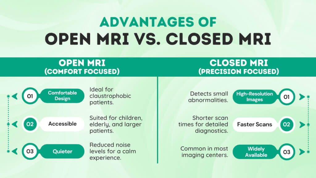
Open MRI is usually better for comfort, claustrophobia, and larger body size, while closed MRI is usually better when image quality and exam flexibility matter most. Closed MRI is usually the right choice when the exam requires the highest image detail, because closed scanners typically operate at higher field strengths (about 1.5T to 3T). Open MRI is usually the right choice when comfort, accessibility for larger patients, or claustrophobia is the limiting factor, because open scanners trade space for lower field strength (about 0.3T to 1.2T).
Wide-bore MRI is the common middle option; it maintains a closed design while offering a larger tunnel and high field strength (often 1.5T to 3T).
So, open MRI vs closed MRI: which can accurately answer your complex condition? This article will explore the differences between the MRI systems, including their advantages and challenges.

Open MRI vs Closed MRI Differences
Open MRI is generally better for comfort and accessibility, while closed MRI is usually preferred for higher-detail imaging and more demanding exams. Check the table for more detailed differentiation.
| Factor | Open MRI | Closed MRI |
|---|---|---|
| Opening design | Open on the sides, with more visible space around the patient. | Tube-like or bore-style design, with the patient positioned inside a more enclosed magnet. |
| Image quality | Can provide high-quality images for many exams, especially with newer systems, but older open MRI units may produce lower image quality and some exams may not be suitable for open MRI. | Generally provides the highest image quality and is typically preferred when maximum detail is needed. |
| Scan speed | Often perceived as less ideal when speed and high-detail imaging are both priorities; exact scan time depends on the machine and exam type. | Commonly preferred for more demanding exams because standard closed-bore systems generally offer stronger imaging performance, which often supports more efficient high-detail studies. |
| Claustrophobia tolerance | Better for patients with claustrophobia because the sides are open and the setup feels less confined. | More difficult for claustrophobic patients because the exam is performed inside a narrow enclosed space. Wide-bore and short-bore systems can help somewhat. |
| Body size / accessibility | Often better for larger patients and for those who need a less confining setup. | Standard closed systems can feel tighter, though newer wide-bore systems may improve comfort for larger patients. |
| Typical use cases | Often chosen for patients with claustrophobia, larger body habitus, or when comfort/accessibility is a major concern. Suitable for many routine exams, but not all. | Usually chosen when the exam requires the best image detail or when specific exam types are better performed on closed systems. |
What are MRI Techniques?
Magnetic Resonance Imaging, also known as MRI, is a powerful diagnostic tool that uses strong magnetic fields, radio waves, and advanced computer technology to create detailed images of the body.
It exploits the properties of hydrogen atoms in your body’s water to create images of soft tissues, such as the brain, muscles, and joints.
The heart of MRI techniques is a powerful magnet, ranging from 0.5 to 3.0 Tesla, that generates the magnetic field. It aligns the hydrogen protons within your body in one direction, like a compass needle pointing toward the Earth’s magnetic field.
The MRI machines then emit short bursts of radiofrequency energy that disturb the proton alignments, temporarily knocking the protons out of their original positions.
When the pulse stops, the protons return to their original positions and realign with the magnetic field. Relaxed protons emit energy signals that the MRI machine detects. The computer then processes these signals and converts them into cross-sectional images.
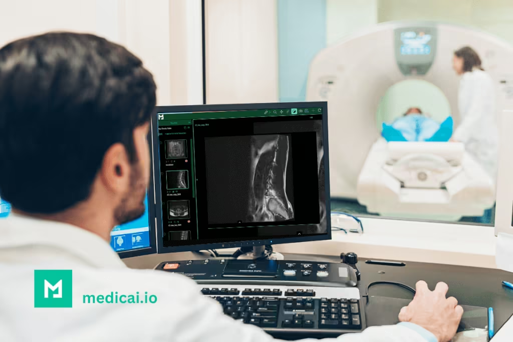
Medicai makes the imaging process a breeze by allowing real-time data sharing and secure cloud PACS storage. Clinicians can quickly access imaging results, enabling them to make faster, smarter decisions.
MRI is beneficial for
- Neurological diagnosis: brain tumors, strokes, multiple sclerosis, etc
- Musculoskeletal imaging: ligament tears, cartilage damage, spinal abnormalities, etc.
- Oncology monitoring: tumor growth or response to treatment
- Cardiology: heart structure and vascular health
MRI comes in two types, open MRI and closed MRI, each with some distinct features.
What Is An Open MRI?
The open MRI is a patient-centered design with open slides or a wider gap. Its less restrictive design offers a comfortable environment for patients with confined-space issues, such as those in a closed MRI.
Open MRI systems are effective for routine imaging, including-
- Musculoskeletal scans: Examining larger structures like bones and major joints.
- Pediatric imaging: Providing a less scary experience for children.
- Geriatric patients: Accommodating mobility challenges and ensuring easier positioning.
- Obesity or Bariatric needs: Furnishing a comfy solution for larger people.
What Does an Open MRI Look Like?
Open MRI scanners feature open sides or a wide C-shaped structure, providing more space around the patient than traditional closed MRI tunnels. Instead of being completely enclosed, patients lie between two magnetic plates — one above and one below — while the sides remain open.
This design helps reduce anxiety, especially for those with claustrophobia, obesity, or pediatric needs. The open space allows for easier breathing, communication with technicians, and even limited movement during longer scans.

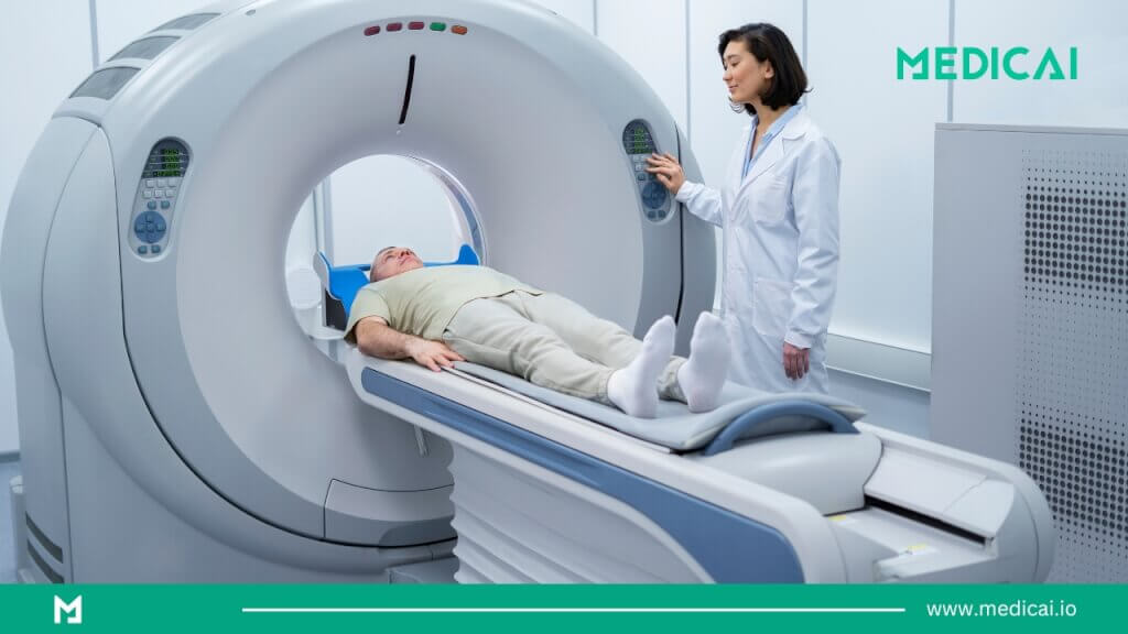
What Is a Closed MRI?
A closed MRI is the traditional design containing a cylindrical tunnel. The patients lie on a table that slides into the machines. The closed MRI design can maximize image quality using strong magnetic fields and advanced imaging technology.
Closed MRI can assess several conditions effectively, including-
- Brain and Spinal Imaging: Detecting minor abnormalities like brain lesions, nerve compression, or herniated discs.
- Cardiac Imaging: Assessing heart structures, blood flow, and vascular abnormalities.
- Orthopedic Scans: joint damage, ligament tears, and cartilage degeneration.
- Oncology: Identifying tumors and monitoring their progression.
What Does a Closed MRI Machine Look Like?
A closed MRI machine resembles a long, cylindrical tunnel (also called a bore). Patients slide into the narrow tube, which contains a powerful magnet that produces high-resolution images.
While the enclosed design may feel intimidating to some, it delivers stronger image quality due to higher magnetic field strength (1.5 T to 3 T).
Open MRI vs Closed MRI: The Key Differences
What do you need – an open MRI or a closed MRI?
To figure it out, you must know how these systems differ in design, functionality, and result. So, let’s check out the differences between open MRI and closed MRI.
Design and Structure
The design of an open MRI is less restrictive, featuring open sides or a wide gap above the patient. The machine is usually C- or U-shaped, creating a more spacious, less confining environment. Thus, you have better visibility during the scan.
However, the magnets in open MRI are a bit weaker. They range from 0.3 to 1.2 Tesla. So, open MRI machine images may have some clarity issues, lacking the fine details seen in closed MRI images.
Closed MRI, on the other hand, produces remarkably detailed images. It features powerful magnets ranging between 1.5 and 3 Tesla. The machine’s cylindrical tunnel fully encloses the patient, which may feel a bit tight and confined.
However, modern closed MRI machines have better ventilation, mirrors, and calming lighting that can help you feel more at ease during imaging.
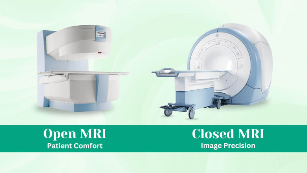
Image Quality
Open MRI has a lower magnetic field strength. So, the images of open MRI machines lack the ultra-high precision that you need for more complex diagnoses. Open MRI is effective for less complex cases, such as scanning for larger injuries or monitoring chronic conditions.
However, even with slightly lower-resolution images, Medicai’s updated platform ensures healthcare providers can make accurate and timely decisions.
On the other hand, closed MRI offers superior image clarity than open MRI images, thanks to its powerful magnets. It produces sharp, precise images required for high-resolution imaging, such as small tumors, brain lesions, cartilage damage, and joint evaluations.
Open MRI vs Closed MRI Image Quality
While both technologies use magnetic resonance, closed MRI scanners generally produce higher-resolution images due to stronger magnets.
However, modern high-field open MRI machines (up to 1.2 T) have significantly narrowed the gap.
| Feature | Closed MRI | Open MRI |
|---|---|---|
| Magnetic Field Strength | 1.5 T – 3 T | 0.3 T – 1.2 T |
| Image Resolution | Excellent – ideal for fine details | Good – suitable for most soft-tissue exams |
| Scan Duration | Faster (15–30 min) | Slightly longer (30–45 min) |
| Comfort | Enclosed; may cause anxiety | Open design; more comfortable |
| Noise Level | Louder | Quieter |
| Accessibility | Limited space | Wheelchair- and pediatric-friendly |
Patient Comfort
MRI can indeed create concerns about comfort. Unlike the CT scan, you may need to remain still for a long time for some scans.
Thanks to its shape, the open MRI machine offers a more relaxed environment. You can see your surroundings during the scan, which comforts and calms you.
Also, the open slides and wide gap are more accessible and comfortable for taller or larger patients, who are especially well-suited to an open MRI.
- Children who get scared in a closed MRI
- Larger patients who may not fit in a closed MRI
- Those with mobility challenges
On the other hand, the enclosed design of a closed MRI, with its narrow tunnel and loud knocking sound, can make you feel confined or uncomfortable. It’s especially difficult for people with claustrophobia, which can increase their anxiety.
However, many facilities now offer noise-canceling headphones or video goggles to distract and comfort patients.
Scan Duration
Open MRI scans take longer to complete because of their weaker magnetic fields. Depending on the complexity, a scan can last 30 to 60 minutes, sometimes more.
On the contrary, closed MRIs are faster, and all credit goes to their strong magnetic fields. A standard scan usually takes 15 to 45 minutes, depending on the diagnosis.
Medicai provides integrated, secure cloud storage with advanced sharing capabilities to preserve the scans. The user-friendly portals make it easy for you and healthcare providers to quickly view and share imaging records.
Is an open MRI accurate enough for my case?
Open MRI can be accurate for many routine exams, but image detail depends on field strength and the clinical question. Open scanners are typically lower-field (about 0.3T to 1.2T), which can reduce fine-detail clarity for subtle findings.
Closed MRI remains the best option for high-detail neuro or vascular studies, and the article already notes that 3T closed MRI is the gold standard for those cases.
Open MRI often works well for musculoskeletal, pediatric, and anxious-patient workflows where comfort improves scan completion and routine diagnostic quality is sufficient.
Cost
Open MRIs cost less than closed MRIs because of their simpler technology and lower magnetic field. Closed MRIs, on the other hand, feature advanced technology, higher magnetic field strength, and superior image quality, which make the imaging expensive.
A closed MRI scan usually costs $1,200 to $4,000, depending on the imaged area and complexity. However, most insurance plans cover closed MRI scans.
Is Open MRI More Expensive?
Open MRI exams can sometimes cost 10 – 20 percent more than closed MRIs because:
- The scanners operate at lower field strengths, requiring longer image acquisition times.
- Fewer facilities have open MRI machines, which may affect scheduling and pricing.
| Type of MRI | Average Cost Range (USD) | Insurance Coverage | Best For |
|---|---|---|---|
| Closed MRI | $1200 – $4,000 | Usually covered | High-resolution brain, spine, or vascular imaging |
| Open MRI | $700 – $5,000 | Covered when medically justified | Claustrophobic or pediatric patients; orthopedic imaging |
Wide-Bore vs Open MRI: What’s the Difference?
Many patients confuse wide-bore MRIs with open MRIs.
- Wide-bore MRI: A larger tunnel (up to 70 cm) within a traditional closed design, offering more space while maintaining high-field strength (1.5 T–3 T).
- Open MRI: Fully open sides with lower magnetic strength (0.3 T–1.2 T).
Both aim to reduce claustrophobia, but wide-bore MRIs deliver closed-MRI-level image clarity with improved comfort.

Advantages and Disadvantages of Open MRI
Let’s look at the pros and cons of picking an open MRI.
Advantages
- It enhances comfort, reduces anxiety, and provides a more relaxed experience for children, larger patients, and people with claustrophobia.
- It offers easy accessibility, especially for patients with special needs or the elderly.
- The reduced noise levels provide a less intimidating environment for noise-sensitive patients.
- Open MRI scans are more affordable.
Disadvantages
- Open MRI images are not high-quality, as the weaker magnetic field may not capture fine details.
- It takes longer, so you have to stay still for a long time.
- Open MRI machines may not be available in all areas.
Advantages and Disadvantages of Closed MRI
Closed MRI comes with both benefits and challenges.
Advantages
- The powerful magnets of a closed MRI machine produce detailed, high-resolution, precise images that can detect small abnormalities, such as tiny tumors or cartilage damage.
- Closed MRI scans take less time because of their powerful magnetic fields.
- Closed MRI machines are widely available in most imaging centers and hospitals.
Disadvantages
- The enclosed tunnel of closed MRI machines can feel overwhelming for patients with claustrophobia or anxiety.
- The machines make loud knocking sounds during scans that can be distressing for many.
- Patients with larger body sizes or mobility limitations may not fit comfortably in a closed MRI machine.
- Closed MRI scans are expensive due to their advanced technology and higher magnetic field strength.
Open MRI vs Closed MRI: Choosing the Right MRI for Your Needs
Open MRI or Closed MRI – the decision usually depends on your medical conditions, complexity, and comfort.
Best default rule:
- Claustrophobia, mobility limits, bariatric needs, pediatric comfort: open MRI or wide-bore MRI, based on the clinical question and scanner field strength.
- Complex brain, spine, vascular, small lesion questions: closed MRI, often 3T when available.
When To Choose Open MRI
Open MRI prioritizes comfort. So, it is ideal for
- Claustrophobic patients
- Children
- Larger people
- People with mobility challenges
However, an open MRI may not provide the higher detail and resolution as a closed MRI. It is effective for general imaging, such as monitoring chronic conditions or assessing larger abnormalities that don’t require ultra-high precision.
When To Choose a Closed MRI
A closed MRI is ideal when you need the highest level of detail, resolution, and precision.
- Brain and neurological disorders
- Spinal conditions
- Cardiac imaging
- Orthopedic Injuries
Whether you undergo an open or closed MRI, Medicai ensures your images and reports are securely stored and accessible in real time. This eliminates delays, allowing your doctor to promptly review and discuss the findings.
Open MRI for Brain and Spine Imaging
Open MRI systems can successfully image the brain, spine, and joints, though they may not capture subtle lesions or microvascular structures as precisely as closed MRI systems.
For high-detail neuro or vascular studies, 3 T closed MRI scanners remain the gold standard.
For musculoskeletal, pediatric, or anxious patients, open MRIs offer a better experience without compromising routine diagnostic quality.
Next step: what to ask when scheduling your MRI
The right MRI choice depends on two constraints: the image detail required for the diagnosis and patient comfort and access constraints.
Scheduling checklist
- Clinical question: brain and vascular details, spine nerves, or small-lesion evaluation usually require a closed MRI with high image quality.
- Field strength: open MRI field strengths are typically lower than those of closed MRI, so the exact Tesla level matters.
- Wide-bore option: wide-bore MRI can reduce claustrophobia while keeping high-field strength.
- Scan duration tolerance: open MRI can take longer; staying still matters for image quality.
- Fit and positioning: Bariatric needs and mobility limits often push the decision toward open MRI or wide-bore MRI.
- Results delivery: image access and report sharing should be clear before the appointment, especially when multiple clinicians need the study.
Imaging record access, after the scan
MRI value depends on the report and images reaching the next clinician fast. A cloud PACS workflow, such as Medicai, can reduce delays by keeping studies accessible for secure sharing and follow-up review, instead of relying on discs or manual transfers.


Related Articles


Lets get in touch!
Learn more about how Medicai can help you strengthen your practice and improve your patients’ experience. Ready to start your Journey?
Book A Free Demo