Adding medical imaging to healthcare software and apps: what teams need to plan for

Medical images used to live almost entirely inside radiology. A study was acquired, read on a dedicated workstation, and stored in an on-premise PACS that few other systems could reach.
That model is changing quickly. Referring physicians expect to open images from the EHR. Care teams want them inside telehealth visits and specialist referrals. Patients want to see and share their own scans without carrying a CD from one clinic to the next. AI tools need a steady, reliable feed of studies to analyze.
For product and IT teams, this means imaging is no longer a radiology-only feature. It is becoming a core capability of clinical software and patient apps alike. Getting it right takes more planning than most teams expect.
Why imaging is harder than other health data
Most health data is small and structured: a lab value, a medication, a diagnosis code. Imaging is different in almost every way that matters to a software team.
- Size. A single CT or MRI study can run to hundreds of megabytes, and some multi-series or 3D studies are far larger. That affects storage costs, upload times and how fast a viewer can open a study on a slow connection.
- Format. Images come wrapped in DICOM, a standard that bundles pixel data with detailed metadata about the patient, the device and the acquisition. Handling DICOM correctly is a specialty in itself.
- Workflow. Imaging touches ordering, scheduling, acquisition, reading, reporting and sharing. Each step often runs on a different system, and the study has to move cleanly between all of them.
- Sensitivity. DICOM headers carry identifying information, and some images can reveal identity on their own. Privacy has to be designed in, not added later.
These factors explain why teams that treat imaging like any other file upload tend to run into performance, integration and compliance problems soon after launch.
The foundation: buy the infrastructure, build the experience
The most expensive mistake teams make is trying to build imaging infrastructure from scratch. Storage, DICOM handling, a diagnostic-quality viewer and secure sharing are mature, specialized capabilities. Recreating them rarely adds value and adds years of maintenance.
A more practical approach is to build on an established imaging layer, such as a cloud PACS or a medical imaging API, and focus internal effort on the experience around it. Whichever platform you choose, check that it supports the standards your product will depend on:
- DICOMweb for retrieving, searching and storing studies over standard web protocols, so web and mobile clients can work with images without legacy DICOM networking.
- HL7 and FHIR for linking imaging to orders, reports and patient records in the EHR. The FHIR ImagingStudy resource is increasingly how applications discover which studies exist for a patient.
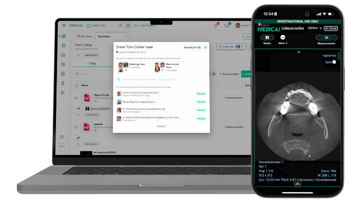
- An embeddable viewer that can open inside your own interface, so users are not sent to a separate application to see a scan.
With that foundation in place, the real product work is deciding how imaging should appear inside each workflow and each app.
Clinical and enterprise software: imaging inside the workflow
For hospitals, imaging centers and specialty groups, the goal is to put images where clinicians already work. Common projects include:
- EHR integration that lets referring physicians open a study from the patient chart with one click, alongside the radiology report.
- Referral and second-opinion portals where outside specialists can review studies securely without accounts on internal systems.
- AI pipelines that route new studies to triage or detection models and return results into the reading worklist.
- Multi-site workflows that let radiologists read across locations as if every study were stored in one place.
These projects sit between systems that were never designed to work together, and each organization’s mix of EHR, RIS, PACS and AI vendors is different. That is why off-the-shelf connectors often cover only part of the need. Many organizations work with a healthcare software development company to build the integration layer, mapping orders, reports and studies across systems while the imaging platform handles storage and viewing.
The key design decision is where the source of truth lives. Keep images in the imaging platform, and let other systems reference them through standard APIs rather than copying files between databases.
Patient-facing apps: making images useful to patients
Patients increasingly expect to access their own imaging the same way they see lab results. For digital health startups and provider organizations, adding imaging to a patient app opens up features such as:
- Viewing scans and reports on a phone or tablet after a visit.
- Sharing a study with a new specialist or for a second opinion in a few taps, instead of requesting a CD.
- Uploading outside imaging before an appointment, so the care team has it ready.
- Seeing images during a telehealth consultation, with the clinician walking through the findings.
These features look simple on screen but are demanding underneath. Large studies must load quickly over mobile networks. Consent and sharing flows must be clear enough for patients to use and strict enough to satisfy compliance teams. The viewer must work well for non-clinical users, who need orientation and context rather than diagnostic tools.
Because mobile performance, privacy and usability all have to come together, teams often bring in a healthcare app development company with both mobile and imaging experience, rather than a general app studio. The best results come from designing with patients early and planning for ongoing releases after launch.
Security, privacy and compliance
Imaging data is protected health information, so every design choice carries compliance weight. Teams should plan for these requirements from the first sprint:
- Regulatory scope. HIPAA applies to US deployments, and GDPR applies when processing data of people in the EU. Many products need to satisfy both.
- Encryption of studies in transit and at rest, including any cached copies on mobile devices.
- Access controls and audit logs that record who viewed, shared or downloaded each study.
- Time-limited, revocable sharing links instead of permanent open access.
- De-identification for research, AI training or teaching use, covering DICOM header fields and any identifying details burned into the pixels.
It is far cheaper to build these controls into the architecture than to retrofit them after an audit or a security review.
A planning checklist before you build
Before committing to a roadmap, product and IT leaders should be able to answer these questions:
- Which users need images, and at which moment in their workflow?
- Which imaging platform will be the source of truth, and does it support DICOMweb, FHIR and an embeddable viewer?
- Which systems need to connect to it, such as the EHR, RIS, AI tools or partner portals?
- How will large studies perform on mobile networks and older devices?
- How will patients give consent, share studies and revoke access?
- Who will maintain the integrations and apps after launch as standards and regulations change?
Conclusion
Imaging is moving out of the reading room and into every corner of care, from the EHR to the patient’s phone. The teams that succeed build on proven imaging infrastructure, invest their own effort in the workflows and experiences around it, and treat security and interoperability as requirements from day one rather than features to add later.

Related Articles

Lets get in touch!
Learn more about how Medicai can help you strengthen your practice and improve your patients’ experience. Ready to start your Journey?
Book A Free Demo