How Brain MRI Segmentation is Revolutionizing Neuroimaging

The human brain is one of the most complex structures in nature, and understanding it starts with seeing it clearly.
Enter brain MRI segmentation: a powerful technique that transforms raw medical scans into labeled maps of brain tissue and pathology. It’s the process of dividing an MRI scan into different regions, like gray matter, white matter, cerebrospinal fluid, or abnormal lesions.
Discover how brain MRI segmentation works, explore the evolution from manual methods to AI-driven tools, and highlight the most promising innovations today.

What Is Brain MRI Segmentation?
Brain MRI segmentation is the process of dividing an MRI scan of the brain into distinct, meaningful regions. These regions could represent types of tissue, anatomical structures, or pathological areas like tumors. The goal is to convert grayscale images into labeled maps that doctors, researchers, or AI systems can analyze more effectively.
In practice, segmentation is a crucial first step in many neuroimaging tasks, as it enables clinicians to measure brain volume, monitor disease progression, plan surgeries, and track treatment responses.
For researchers, it opens the door to studying brain structure, aging, and neurological development in large populations.
Types of Brain MRI Segmentation
Depending on the use case, brain MRI segmentation falls into one of three main categories.
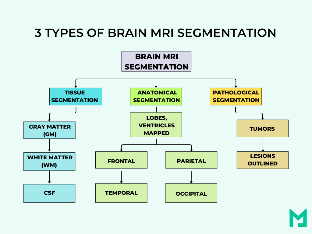
Tissue Segmentation
This type breaks down the brain into its three core tissue types:
- Gray Matter (GM): Where neurons are densely packed; critical for processing information.
- White Matter (WM): The brain’s communication network, made of nerve fibers.
- Cerebrospinal Fluid (CSF): The protective fluid surrounding the brain and spinal cord.
Tissue segmentation is widely used in studies of brain development, aging, and disorders like Alzheimer’s disease and multiple sclerosis.
Pathological Segmentation
It focuses on identifying abnormal areas within the brain, such as:
- Brain tumors
- Edema (swelling)
- Lesions caused by stroke or multiple sclerosis
Accurate pathological segmentation helps with early diagnosis, surgical planning, and monitoring of disease progression.
Anatomical Segmentation
This approach divides the brain into specific regions and substructures, such as:
- Lobes (frontal, temporal, parietal, occipital)
- Cortical areas
- Ventricles
- Deep gray nuclei (e.g., thalamus, putamen)
Anatomical segmentation is vital for surgical navigation and mapping brain function.

The Technical Foundations: How Brain MRI Segmentation Works
Brain MRI segmentation begins with understanding data dimensions and limitations. Poor input can hinder even the best models, so a solid technical foundation is crucial.
Image Characteristics
Brain MRI data can come in two forms:
- 2D slices: single-plane images, commonly used in clinical routines.
- 3D volumes: full brain scans composed of stacked slices across multiple planes (axial, sagittal, coronal).
While 2D segmentation is faster and requires less computational power, 3D segmentation provides richer context. However, 3D data also introduces challenges: more memory use, longer training times, and the need for consistent preprocessing across slices.
Challenges That Affect Segmentation
Common MRI artifacts that affect segmentation include-
- Noise: Random variation in pixel intensity—often caused by low signal strength.
- Partial Volume Effect (PVE): When a voxel contains more than one tissue type, it blurs the boundaries.
- Bias Field (Intensity Inhomogeneity): A gradual shading artifact that makes the same tissue appear brighter in one region and darker in another.
These issues can confuse even the best models. That’s why preprocessing is non-negotiable.
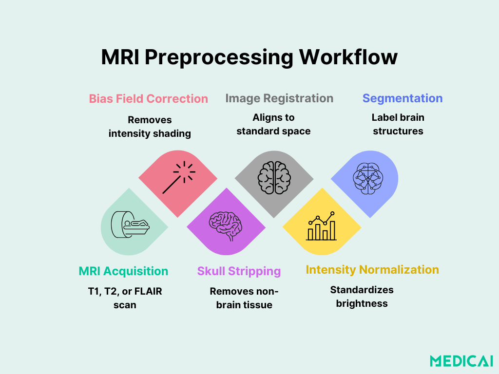
Preprocessing Workflow
Without proper preparation, segmentation results can be inconsistent or completely unusable. Preprocessing guarantees that scans are clean, aligned, and ready for analysis.
Let’s look at a simplified preprocessing pipeline commonly used before segmentation in research settings.
- Image Acquisition – Raw MRI data is collected, usually in T1-weighted, T2-weighted, or FLAIR sequences.
- Bias Field Correction – Intensity inhomogeneity is corrected using tools like N4ITK (Nonparametric Nonuniform Intensity Normalization).
- Skull Stripping (Brain Extraction) – The brain is isolated using algorithms like BET (Brain Extraction Tool in FSL) or SPM’s tissue masks.
- Image Registration – Align the scan to a standard space (e.g., MNI152) using SPM, FSL FLIRT, or ANTs.
- Intensity Normalization – Pixel intensities are standardized across subjects or sessions to reduce variability.
Traditional Segmentation Techniques
Before deep learning, brain MRI segmentation relied on classical image processing techniques. They laid the groundwork for modern methods and remain useful in certain contexts.
Manual Segmentation
Manual segmentation involves experts outlining brain structures slice by slice. It’s the gold standard for creating training datasets and validating new methods.
But there’s a catch!
Manual work is slow, prone to human variability, and not scalable for large datasets. A single scan can take hours to annotate properly.
That’s why automation in MRI segmentation became a top priority.
Thresholding and Region Growing
Thresholding
One of the simplest methods. You pick an intensity value (the threshold), and everything above or below it gets grouped into a segment.
It works well when contrast is high (e.g., CSF vs. tissue). However, the process fails when intensities overlap or when images have noise or artifacts.
Region Growing
Start with a “seed point” (a known tissue pixel), then “grow” the region by adding neighboring pixels with similar intensity.
This technique is good for detecting connected regions (like large lesions). However, it’s highly sensitive to seed placement and noise.
Clustering Techniques
These methods treat each pixel or voxel like a data point in a feature space (based on intensity and sometimes location). Then they group similar data points together.
K-Means Clustering
- Unsupervised
- Classifies data into k groups based on intensity
- Fast and simple, but assumes uniform class shapes
Fuzzy C-Means (FCM)
- Allows partial membership
- Better at handling partial volume effects than hard clustering
Atlas-Based Segmentation
In this method, a pre-labeled probabilistic brain atlas is registered to a patient’s scan. The labeled regions are then transferred over.
The technique is great for anatomical segmentation. However, it struggles with deformed brains and relies heavily on accurate registration.
Validation Techniques
These older methods were validated using manually labeled data or synthetic datasets like:
- BrainWeb: simulated brain MRIs with known ground truth
- IBSR: real human MRI scans with expert annotations
Metrics like Dice coefficient, Jaccard index, and Pixel Accuracy were used to judge performance, and these are still used today.
Deep Learning in Brain MRI Segmentation: A Game Changer
Deep learning revolutionized brain MRI analysis, with CNNs outperforming traditional methods in speed and accuracy.
Deep learning doesn’t need explicit rules. It learns patterns directly from data. That’s incredibly powerful in brain MRI, where structures vary from person to person and even scan to scan.
Do you know why it’s a breakthrough?
- No manual feature engineering—the network figures out what matters.
- Handles complex textures, like tumor borders or gray/white matter overlaps.
- Learns context and distinguishes tissue types based on location and structure.
Unlike classical methods, which struggle with noisy or deformed scans, deep models can be trained to “expect” variability and still perform well.
U-Net: The Workhorse of Medical Image Segmentation
If there’s one model that’s changed the game, it’s U-Net.
Originally developed in 2015 for biomedical segmentation, U-Net is a fully convolutional network with a symmetrical “U-shaped” structure:
- The encoder captures features from the image.
- The decoder reconstructs the segmented output, using skip connections to retain fine-grained details.
U-Net shines in medical settings because:
- It works well with small datasets.
- It produces pixel-perfect segmentation maps.
- It can be adapted for 2D or 3D inputs.
A lightweight U-Net model using 2D slices from multiple planes (sagittal, coronal, transverse) achieved up to 89% mean IoU without data augmentation—fast, efficient, and surprisingly accurate.
Real-World Applications of Brain MRI Segmentation
Brain MRI segmentation is actively applied in hospitals, research labs, and medical imaging startups to address significant issues.
Tumor Segmentation
Brain MRI segmentation is used in identifying and outlining brain tumors (like gliomas) for diagnosis, surgical planning, or treatment monitoring.
In this task, precision matters, especially around tumor boundaries, surrounding edema, and necrotic core regions. Deep learning models, especially U-Net variants, have significantly improved segmentation accuracy in datasets like BRATS and BITE.
Tissue & Structural Segmentation
It’s used in classifying core brain tissues—gray matter (GM), white matter (WM), and cerebrospinal fluid (CSF)—and dividing the brain into anatomical regions.
It is crucial for:
- Studying neurodevelopment in children
- Tracking brain atrophy in aging or Alzheimer’s
- Monitoring lesions in conditions like multiple sclerosis
Most models here are trained on T1-weighted MRI and sometimes utilize multi-modal inputs. Tools like FreeSurfer, FSL FAST, and newer models like QuickNAT and SynthSeg offer fast and reliable outputs suitable for both research and clinical pipelines.
Anatomical Segmentation (Lobes, Ventricles, Subcortical Structures)
Brain MRI segmentation helps in mapping out precise regions of the brain, such as:
- Frontal, parietal, temporal, and occipital lobes
- Lateral and third ventricles
- Hippocampus, amygdala, thalamus
This level of segmentation helps:
- Neurosurgeons plan interventions
- Researchers study functional connectivity
- Psychiatrists assess structural biomarkers in disorders like schizophrenia or depression
Medicai bridges the gap between cutting-edge segmentation research and real-world clinical practice. Our advanced AI models with seamless clinical integration deliver fast, accurate MRI brain image segmentation across diverse real-world settings.
Conclusion
Brain MRI segmentation has come a long way—from manual outlines to real-time AI-powered precision. With deep learning models, what once took hours can now be done in seconds, with greater accuracy and consistency.
Medicai delivers segmentation results that are ready for clinical review across different MRI protocols, patient populations, and institutions.


Related Articles


Lets get in touch!
Learn more about how Medicai can help you strengthen your practice and improve your patients’ experience. Ready to start your Journey?
Book A Free Demo