
Computed tomography (CT) scans are a cornerstone of medical imaging, helping doctors diagnose various conditions. But what if there was a way to make these scans even more accurate and detailed? Enter photon-counting CT (PCCT).
This newer technology promises to revolutionize CT imaging, offering several advantages over traditional energy-integrating CT (EICT). This post will explore the key differences between PCCT and EICT, focusing on their impact on liver lesion detection.
Differences between Photon Counting CT vs. Energy-Integrating CT
| Dimension | Photon-counting CT (PCCT) | Energy-integrating CT (EID-CT) |
|---|---|---|
| Detection principle | Counts individual photons and assigns to energy bins | Integrates total energy over time |
| Conversion | Mostly direct conversion semiconductor | Mostly indirect (scintillator → photodiode) |
| Spectral imaging | “Always on” spectral capability (multi-bin) | Spectral requires dual-energy or special hardware modes |
| Noise and dose efficiency | Better electronic noise suppression, often improved dose efficiency | Strong baseline performance, electronic noise matters more at low signal |
| Spatial resolution potential | Often higher (smaller detector elements) | Limited by scintillator optics and septa geometry |
| Key pain points | Pile-up, charge sharing, spectral corrections | Fewer spectral degrees of freedom unless dual-energy |
How Does Traditional CT (EICT) Work?
Traditional CT scanners, also known as energy-integrating CT (EICT), measure the total energy of X-rays passing through the body. This data then creates detailed images of internal organs, bones, and tissues.
What is a Photon Counting CT Scan?
Photon-counting CT (PCCT) counts individual photons and measures their energy levels, unlike conventional CT, which measures total energy. This allows for higher-resolution images with less noise, improved contrast between tissues, and the potential for lower radiation doses.
PCCT’s ability to differentiate materials based on their energy absorption opens doors for advanced applications in various medical fields, leading to earlier and more accurate diagnoses.
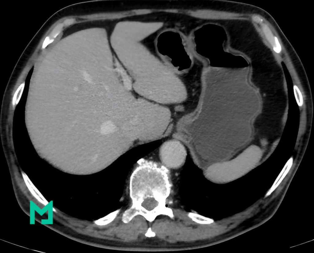
Photon-counting CT vs energy-integrating CT for liver lesion detection
Photon-counting CT improves liver lesion detectability compared with energy-integrating CT, with the greatest gains reported for small and low-contrast lesions when low-keV spectral reconstructions are used.
What the best comparative evidence says
A 2024 virtual framework study reported objective improvement in liver lesion detection and image-quality metrics for PCCT compared with EID-CT, with stronger benefits in more challenging conditions such as lower contrast lesions, lower dose, and larger patients.
Another study using virtual imaging reported improved sensitivity for detecting small liver lesions under 1.5 cm with photon-counting CT compared with energy-integrating detector CT.
A 2024 study on photon-counting detector CT virtual monoenergetic images focused on liver lesion detection showed that “optimal” keV for detectability changes with phantom size and dose, which matters because protocol defaults can underperform in larger patients.
A separate low-energy VMI comparison found improved detectability and image quality for low-keV VMIs on a photon-counting CT system compared with an energy-integrating dual-source CT comparator, supporting the real mechanism behind “lesion pops more at low keV.”
Why PCCT tends to help liver lesion detection
Photon-counting CT improves liver lesion detection through 3 mechanisms that directly address hepatic imaging pain points.
- Noise behavior at low signal: Photon-counting detector CT reduces electronic noise and improves low-keV VMI usability, raising low-contrast lesion conspicuity in the portal venous phase, where differences can be subtle.
- Spectral leverage without “special mode” dependency: Photon-counting CT provides energy-resolved data as a standard output, which makes low-keV VMIs and iodine-related post-processing more available as a routine reading set.
- Spatial resolution potential for small lesions: Photon-counting CT supports smaller detector elements and higher spatial resolution, which improves edge definition and small-lesion visibility, especially when slice thickness and reconstruction parameters are tuned for detection.
Protocol moves that improve liver lesion detection on PCCT
Photon-counting CT performance depends on reconstruction choices, not scanner labels. A PCCT liver lesion detection set works best when the reading stack forces a quick “confirm in 2 series” habit.
Reading stack for liver lesion detection
- Blended or standard series, baseline confirmation, and stability check.
- Low-keV VMI series: lesion conspicuity pass; typically, the series in which hypoattenuating lesions separate better from parenchyma.
- Iodine-focused series when available, characterization support for enhancement differences, especially for small lesions.
KeV selection rule that prevents false confidence
Photon-counting detector CT VMI detectability shifts with patient size and dose, so one fixed keV value is not a universal best setting.
A practical workflow uses low-keV for detection, then a higher-keV or blended series for confirmation when noise texture or beam-hardening confounds appear.
Dose and size reality check
Virtual framework work reported the greatest PCCT advantages in more challenging cases, such as low-dose and larger patients, so protocol validation needs “large habitus, low contrast” test cases, not only ideal conditions.
When EID-CT closes the gap
Energy-integrating CT narrows the difference when the EID-CT workflow already runs optimized dual-energy liver protocols with carefully chosen monoenergetic reconstructions and modern denoising or iterative reconstruction.
Energy-integrating CT still requires selecting spectral modes up front, whereas photon-counting CT provides spectral data by default, which affects the reliability of “spectral rescue” in routine scheduling.
Limits and risks to state explicitly
Photon-counting CT liver lesion evidence still includes a meaningful share of virtual and phantom-based validation, so clinical outcome claims need restraint even when detectability metrics improve.
Photon-counting CT introduces spectral-specific correction issues, such as pile-up and charge sharing, that can affect low-keV appearance and quantitative outputs, making local protocol QA non-negotiable.
What Makes Photon Counting CT (PCCT) Different?
PCCT utilizes a fundamentally different approach. Instead of measuring the total energy, it counts individual X-ray photons and records their energy level. This allows for:
- Improved Image Quality: PCCT produces images with less noise and higher resolution, making spotting small or subtle abnormalities easier.
- Reduced Radiation Dose: PCCT can achieve the same image quality with a lower radiation dose than EICT.
- Enhanced Contrast: PCCT can more effectively differentiate between different materials, leading to better contrast between healthy and diseased tissues.
PCCT and Liver Lesion Detection
A recent study published in The Journal of Medical Imaging explored the benefits of PCCT for liver lesion detection. Researchers used computer simulations to compare PCCT and EICT scans of livers with various lesions. The results were impressive:
- Superior Lesion Detection: PCCT consistently outperformed EICT in detecting liver lesions, especially small or low-contrast lesions.
- Enhanced Image Quality: PCCT images showed improved contrast-to-noise ratio and overall image quality, making it easier for radiologists to identify and characterize lesions.
- Dose Efficiency: PCCT achieved better lesion detection even at lower radiation doses, potentially minimizing patient risks.
PCCT vs EICT: What This Means for Patients
The improved lesion detection and image quality offered by PCCT could translate into:
- Earlier Diagnosis: Detecting liver lesions earlier, when they are more minor and potentially more treatable.
- More Accurate Diagnosis: Reducing the number of false positives and negatives, leading to more confident diagnoses.
- Personalized Treatment: Providing more detailed information about the size, shape, and composition of lesions, enabling more tailored treatment plans.
The Future of CT Imaging
While PCCT is still relatively new, early research suggests it could significantly improve CT imaging, particularly for liver lesions and lung cancer detection. As PCCT becomes more widely available, it could lead to more accurate diagnoses, earlier interventions, and better patient outcomes.


