Cardiology PACS vs. Radiology PACS: Why Specialized CVIS Integration Matters

Precision is the baseline in modern healthcare, but efficiency is the differentiator.
While many institutions attempt to shoehorn cardiac imaging into general Radiology systems, the reality is distinct: The heart is dynamic, and your imaging system must be too.
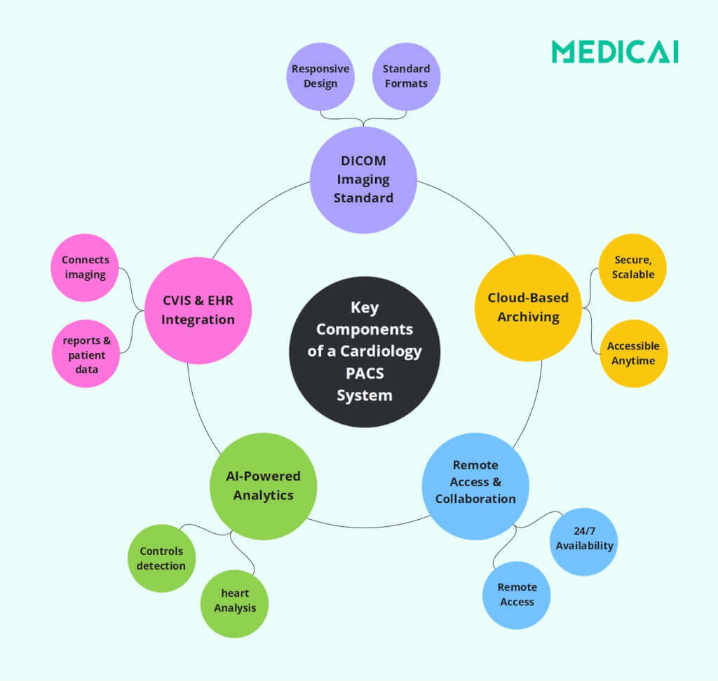
A dedicated Cardiology PACS (Picture Archiving and Communication System) is not just a storage locker for images. It is the operational backbone of the Cath Lab, Echocardiography, and Vascular departments. It must bridge the gap between dynamic imaging (CINE loops) and physiological data (Hemodynamics) to create a unified patient record.
This guide explores why a specialized Cardiovascular Information System (CVIS) integration is critical for reducing physician burnout, improving report turnaround times, and ensuring data integrity.
Why a Standard Radiology PACS Can’t Handle the Heart
The fundamental difference between Radiology and Cardiology is motion. Radiology primarily deals with static images (CR, DR, CT), while Cardiology primarily deals with high-frame-rate video.
If your facility relies on a generic enterprise viewer, you have likely encountered the “stutter” effect—where diagnostic quality is lost during playback. A dedicated Cardiology PACS is engineered with specific attributes to handle these unique demands:
- Dynamic Visualization: Tools for Multi-Planar Reconstruction (MPR) and 3D rendering are essential for planning complex interventions, such as TAVR (Transcatheter Aortic Valve Replacement).
- CINE Loop Playback: Unlike static X-rays, angiograms, and echocardiograms require smooth, high-definition video playback. A specialized PACS ensures multi-frame DICOM files play instantly without buffering or compression artifacts.
- Multi-Modality Harmonization: A single patient often traverses the Cath Lab, Echo, IVUS (Intravascular Ultrasound), and ECG. A true Cardiology PACS creates a “patient-centric” view that aggregates these diverse file types—from waveforms to video—into a single timeline.
The Ecosystem: Linking PACS, CVIS, and Hemodynamics
A PACS never exists in a vacuum. It exists within a vector of related systems. The most critical relationship is between the PACS and the Hemodynamic Monitoring System.
The “Discrete Data” Advantage
Legacy workflows treat images and data as separate islands. The images live in the PACS, and the pressures/gradients live in the Hemodynamic system. This forces Interventional Cardiologists to toggle between screens to understand the full clinical picture.
Modern CVIS integration solves this by ingesting discrete data.
The Result: When a physician opens the study in the PACS viewer, they see the physiological waveforms overlaying the anatomical images. This context is vital for accurate diagnosis in complex vascular cases.
How it works: The PACS/CVIS pulls distinct data points (e.g., ventricular pressures, valve gradients) directly from the Hemodynamic cart.
DICOM Structured Reporting (SR): Eliminating the “Swivel-Chair”
One of the most significant contributors to physician burnout and transcription errors is the “swivel-chair” workflow.
The Old Way: A sonographer performs an echo, freezes the image, measures the Ejection Fraction (EF), and then manually types those numbers into a separate reporting station. The Risk: Manual entry is slow and prone to typos. A decimal point error in a valve area measurement can change a diagnosis.
The Modern Solution: DICOM Structured Reporting (SR) A semantic-aware Cardiology PACS utilizes DICOM SR to automate this pipeline:
- Capture: The sonographer measures on the cart.
- Transfer: The measurements are sent as structured data (not just pixel text) to the PACS.
- Auto-Population: The system automatically maps these values into the final report template.
This workflow allows the reading physician to validate data rather than dictate it, significantly reducing Report Turnaround Time (TAT).
Beyond Storage: AI-Driven Diagnostics and Triage
A modern Cardiology PACS is no longer just a passive archive; it is an active diagnostic partner. While legacy systems rely entirely on manual interpretation, advanced CPACS solutions now integrate Artificial Intelligence (AI) algorithms directly into the workflow.
- Predictive Analytics: By analyzing historical data in the CVIS, the system can identify patterns in patient history that indicate higher risk, aiding proactive care management.
- Automated Quantification: AI algorithms can now automatically calculate Ejection Fraction (EF) and Global Longitudinal Strain (GLS) from echocardiograms, presenting them to the cardiologist for validation. This significantly speeds up reading time.
- Intelligent Triage: AI can analyze incoming CT Angiograms for signs of Pulmonary Embolism (PE) or aortic dissection, flagging critical cases to the top of the worklist for immediate review.
Zero-Footprint Access and VNA Architecture
For PACS Administrators and IT Directors, the “Attribute” that matters most is deployment. The days of installing heavy client software on every workstation are over.
The Zero-Footprint Viewer
Modern architecture demands an HTML5 Zero-Footprint Viewer. This allows cardiologists to access diagnostic-quality images, including CINE loops, via a standard web browser (Chrome, Edge) on any device.
- Vectors: This is critical for Teleradiology, remote on-call consults, and bedside rounding on tablets.
Vendor Neutral Archive (VNA) Strategy
Data migration is a massive pain point. Proprietary PACS often lock images into “wrappers” that make future migration expensive.
The Solution: Adopting a VNA (Vendor Neutral Archive) approach ensures that your cardiac data is stored in a standard, non-proprietary format. This decouples the storage layer from the application layer, giving you the freedom to switch PACS vendors in the future without a painful data migration project.

Deployment Models: Cloud, On-Premise, or Hybrid?
One of the most critical architectural decisions for a healthcare organization is the deployment model. The choice impacts security, speed, and budget (CapEx vs. OpEx).
The Sweet Spot: Many modern facilities utilize a hybrid model—keeping a local cache (Edge server) for instant image retrieval in the Cath Lab, while syncing to the cloud for long-term archiving and remote access.
Cloud-Native PACS (SaaS)
- Best for: Scalability and multi-site networks.
- Advantage: Reduces IT maintenance overhead and shifts costs to an operational model (OpEx). It offers built-in Disaster Recovery (DR) and allows for rapid scaling without buying new hardware.
On-Premise
- Best for: Facilities with limited internet bandwidth or strict data sovereignty requirements.
- Advantage: fast, local access to heavy data files (like CINE loops) regardless of internet connection status.
Hybrid Architecture
- The Sweet Spot: Many modern facilities utilize a hybrid model—keeping a local cache (Edge server) for instant image retrieval in the Cath Lab, while syncing to the cloud for long-term archiving and remote access.
Checklist: How to Choose the Right Cardiology PACS
When evaluating vendors, move beyond the brochure. Use this checklist to verify they support the essential Attributes of a modern cardiac workflow:
- DICOM SR Support: Does it auto-populate report fields from Echo and Cath carts?
- Hemodynamic Integration: Can it import waveforms and pressure gradients as discrete data?
- Zero-Footprint Viewer: Can Interventional Cardiologists view diagnostic-quality angiograms from home?
- VNA Compatibility: Is the storage architecture truly vendor-neutral?
- HL7 FHIR Readiness: Is the system prepared for modern EMR interoperability (e.g., Epic/Cerner)?
- Single Sign-On (SSO): Does it integrate with your hospital’s Active Directory for seamless security?

Conclusion
Cardiology is a discipline of motion, pressure, and flow. A generic imaging platform simply cannot capture the nuance required for high-level cardiac care. By investing in a Cardiology PACS that prioritizes CVIS integration, Hemodynamic data, and Structured Reporting, healthcare providers don’t just get software—they get a workflow engine that improves data integrity and gives their physicians time back.


Related Articles
Lets get in touch!
Learn more about how Medicai can help you strengthen your practice and improve your patients’ experience. Ready to start your Journey?
Book A Free Demo