Medical Imaging Technology
The Medical Imaging Technology category highlights the latest advancements and innovations in medical imaging. This section covers topics such as the development of new imaging techniques, improvements in diagnostic accuracy, and the integration of technologies like artificial intelligence and cloud-based solutions. Articles explore how these innovations are transforming the way healthcare professionals capture, analyze, and utilize imaging data to improve patient care and streamline medical workflows.
125 posts
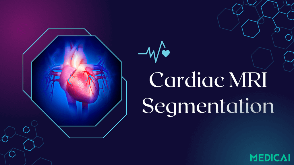
Read MoreFrom Manual to AI: The Future of Cardiac MRI Segmentation
From Manual to AI: The Future of Cardiac MRI Segmentation What if a heart scan could reveal life-saving details in the time it takes to sip your coffee? That’s the promise of modern cardiac MRI segmentation. Cardiac MRI segmentation is the process of dividing scans into the left ventricle, right...

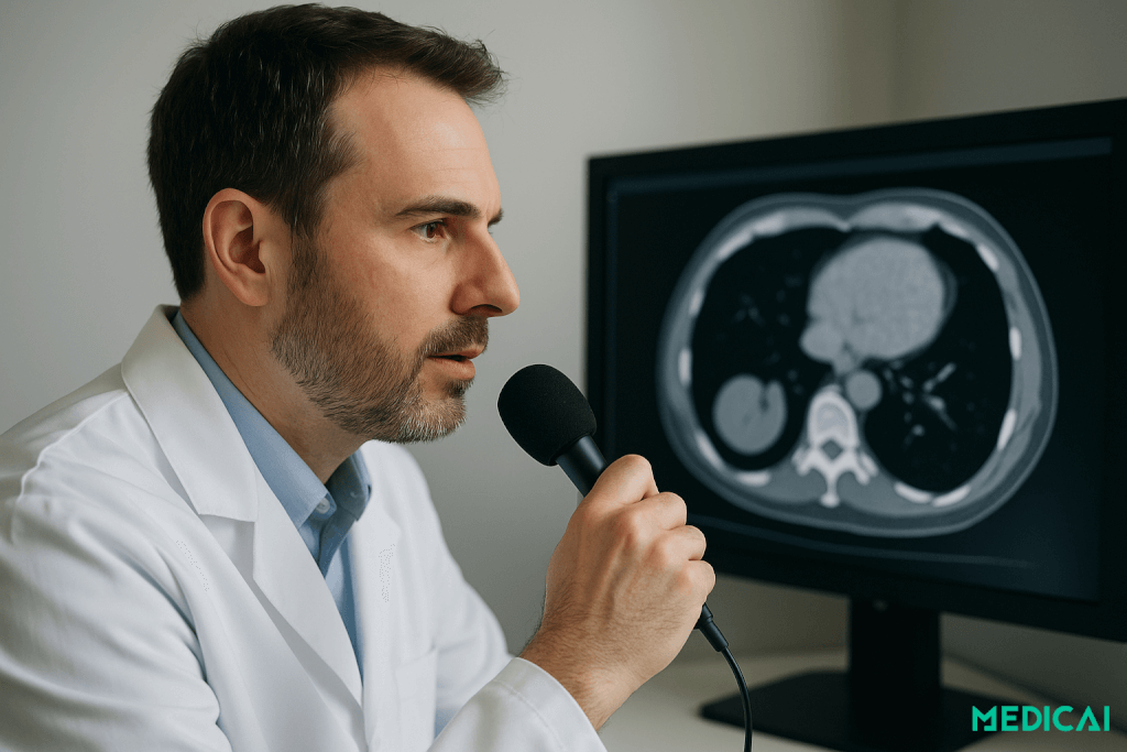
Read MoreVoice‑Enabled Radiology: From Dictation to Contextual Command
Voice‑Enabled Radiology: From Dictation to Contextual Command What if your radiology workstation could not only listen, but also understand? This is the promise of voice-enabled radiology. Voice-enabled radiology is the evolution from traditional dictation to AI-powered contextual commands. Instead of only transcribing words, modern systems understand intent....

Read MoreWhy Structured Annotations Are the Future of Oncologic Reporting?
Why Structured Annotations Are the Future of Oncologic Reporting? Cancer imaging is evolving fast, and so are the expectations. Clinicians need instant clarity, patients deserve precision, and radiologists face growing pressure to deliver both. Structured annotations in oncologic reporting embed standardized data onto PACS images, creating faster, clearer reports...

Read MorePACS for Urgent Care: Setup, Integrations, Best Practices
PACS for Urgent Care: Setup, Integrations, Best Practices In urgent care, every minute matters. Patients want answers now, not after a CD burns or a courier arrives. That’s where PACS comes in. A modern Picture Archiving and Communication System puts every X-ray, CT, and ultrasound in one secure...

Read MorePACS Integration for Modern Veterinary Practices
PACS Integration for Modern Veterinary Practices Tired of chasing down images, repeating data entry, or flipping between systems just to finalize a patient report? Well, here’s a better way to handle your work: Vet PACS integration. Vet PACS integration brings all your critical tools together: imaging,...

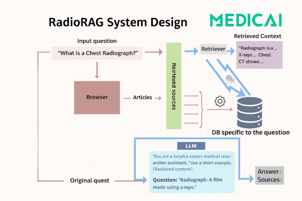
Read MoreRetrieval-Augmented Generation: The Missing Link Between AI and Radiology Accuracy
Retrieval-Augmented Generation: The Missing Link Between AI and Radiology Accuracy As radiologists face mounting caseloads and increasing demand for rapid diagnostic decisions, large language models (LLMs) have emerged as promising allies. Yet, conventional LLMs suffer from limitations that constrain their reliability: hallucinations, outdated information, and a lack of source transparency....

Read More1.5T vs 3T MRI: Which Scanner Fits Your Clinical Needs?
1.5T vs 3T MRI: Which Scanner Fits Your Clinical Needs? Ever wondered why some clinics stick with 1.5T magnets while others upgrade to 3T? 1.5T scanners deliver reliable, implant-safe imaging and predictable scheduling, while 3T systems provide up to double the signal–to–noise ratio for finer detail and faster scans. The...

Read MoreHow to Convert DICOM to JPEG in Minutes
How to Convert DICOM to JPEG in Minutes Fastest way to convert DICOM to JPEG or JPG The DICOM to JPEG process extracts and window-levels the raw pixel data, then compresses it into a standard JPEG file. This process preserves key diagnostic details while making images instantly viewable...

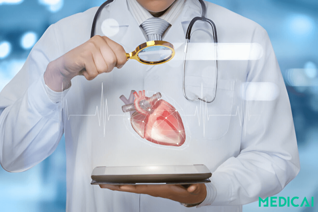
Read MoreContrast MRI: From Prep to Image Clarity
Contrast MRI: From Prep to Image Clarity What if your MRI scan could reveal hidden tumors or tiny vessel leaks in stunning detail? Contrast MRI acts like a spotlight, bringing unseen abnormalities into clear view. MRI with contrast uses intravenous gadolinium chelates to shorten T1 relaxation, making...

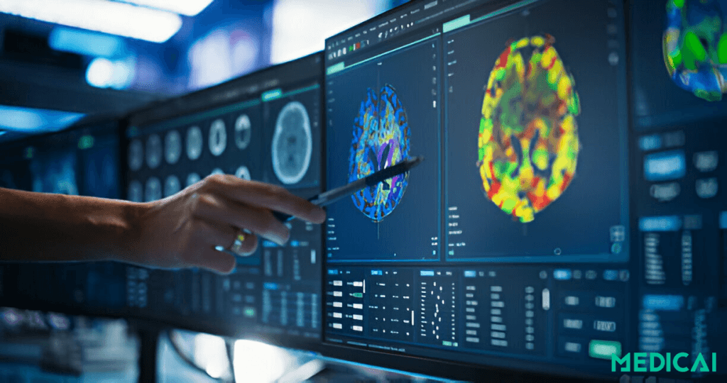
Read MoreHow Deep Learning Revolutionizes Cardiac MRI Segmentation
How Deep Learning Revolutionizes Cardiac MRI Segmentation What if your next cardiac MRI report arrives before you finish your coffee? Fully automatic deep-learning segmentation combines a CNN-powered ROI detector, U-Net–based backbone, and deformable-model refinement to deliver pixel-perfect chamber masks in seconds. The result is clinician-grade contours, Dice...

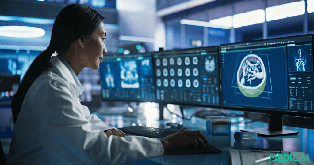
Read MoreFrom Scan to Diagnosis: How Does PACS Works
From Scan to Diagnosis: How Does PACS Works No film, no delays, just a click and your images appear! That’s the power behind modern imaging workflows, such as PACS, where speed and precision transform patient care. PACS (Picture Archiving and Communication System) digitizes film archives, capturing, storing, and...

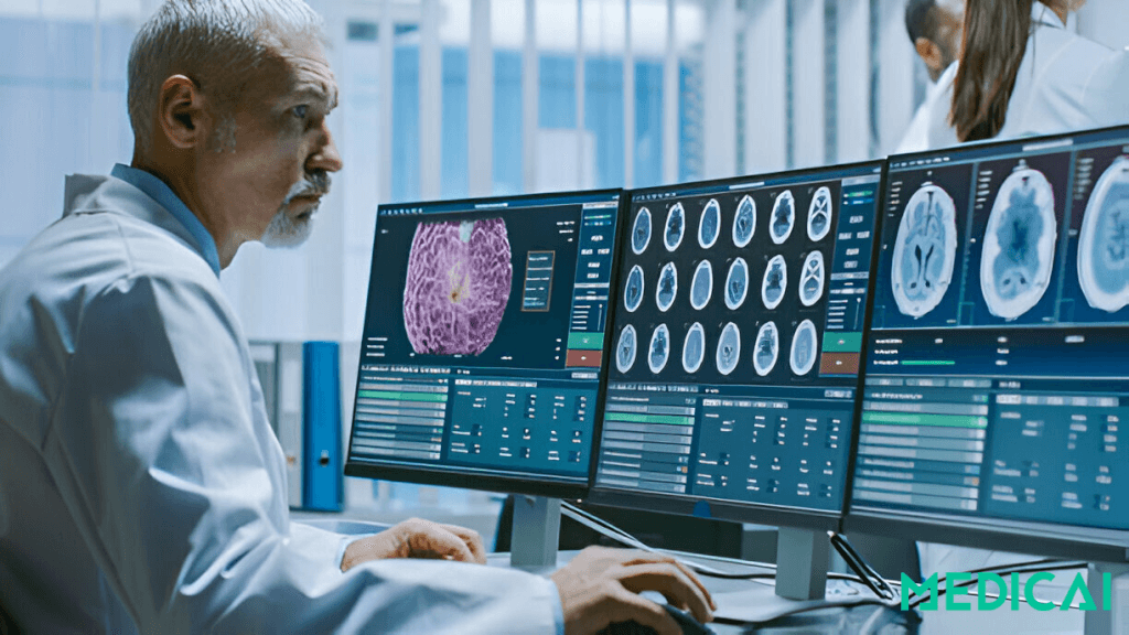
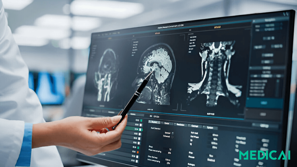
Read MoreBrain Tumor MRI Image Segmentation Using Deep Learning Techniques
Brain Tumor MRI Image Segmentation Using Deep Learning Techniques Imagine reducing hours of manual brain MRI segmentation to seconds. Brain tumor boundaries emerge automatically, and deep learning is making this possible. It brings precision, speed, and consistency to neuro-oncology workflows. Deep learning leverages CNNs, attention U-Nets, and transformer hybrids...
