Interventional Radiology: Safe, Fast & Effective Procedures

What if life-saving treatments no longer required large incisions, long hospital stays, or weeks of painful recovery?
Interventional radiology (IR) is making that a reality.
Interventional radiology (IR) is a medical specialty that uses image-guided, minimally invasive techniques to diagnose and treat diseases. It uses small incisions to reduce pain, recovery time, and risks while effectively treating vascular conditions, cancer, organ disorders, and chronic pain.
Let’s find out if IR is leading the future of precision medicine, offering safer and more effective treatments.

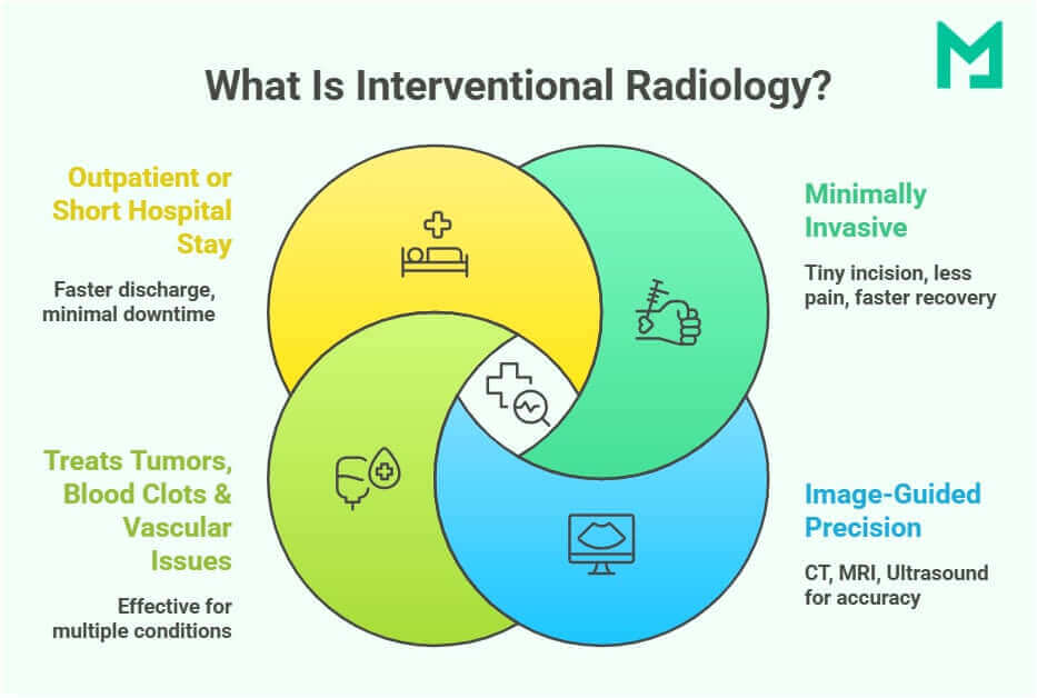
What is Interventional Radiology?
Interventional radiology (IR) is a minimally invasive, image-guided medical specialty that uses advanced imaging techniques, such as X-ray fluoroscopy, ultrasound, CT, and MRI, to diagnose and treat various conditions.
Unlike traditional surgery, IR procedures require only small incisions, leading to less pain, fewer complications, and faster recovery times.
IR is widely used in:
- Vascular interventions
- Oncology
- Musculoskeletal treatments
- Pain management.
Interventional radiology bridges radiology and surgery, improving disease detection and treatment while reducing open surgeries. Over the past century, it has evolved from a diagnostic tool to a therapeutic specialty with specialized procedures.
Modern IR suites combine imaging modalities for greater precision in interventions. These advanced imaging environments provide the following:
- Real-time visualization of complex anatomy for accurate interventions.
- Lower radiation exposure compared to conventional fluoroscopy-based techniques.
- Improved treatment outcomes in areas like stroke management, tumor ablation, and vascular interventions.
Advancements in AI, robotics, and imaging are redefining minimally invasive medicine. Interventional radiology offers safer, faster, and more effective treatments, becoming essential in modern patient care and precision medicine.
Medicai’s imaging solutions greatly enhance interventional radiology (IR). We support IR by improving image clarity, speeding up abnormality detection, and reducing radiation exposure to enhance patient safety.
The Foundations of Interventional Radiology
Interventional radiology (IR) is built on the principles of precision, minimally invasive techniques, and advanced imaging guidance.
Minimally Invasive Approach: A Game-Changer in Medicine
One of the most significant advantages of IR is its minimally invasive nature, which provides a safer alternative to conventional surgeries. Using needle-sized access points, interventional radiologists can diagnose and treat conditions deep within the body without open surgery.
Thus, it offers-
- Shorter Hospital Stays
- Reduced Risk of Complications
- Faster Recovery
- Less Pain and Scarring
IR dramatically improves outcomes for high-risk surgical patients with heart disease, diabetes, or weakened immune systems. It has revolutionized vascular treatments, cancer therapies, and organ management by offering safer alternatives.
Key Imaging Modalities in Interventional Radiology
IR relies on advanced imaging technologies to guide procedures with real-time precision. These imaging modalities allow interventional radiologists to visualize structures inside the body, accurately place instruments, and monitor progress during treatment.
- X-ray (Fluoroscopy): This technique offers continuous X-ray imaging for guiding catheters in procedures like angioplasty and embolization. It also allows interventional radiologists to track the movement of contrast dye for precise vascular interventions.
- Ultrasound: It uses sound waves for dynamic images, making it ideal for biopsies, vascular access, and tumor ablation. It’s also radiation-free, ensuring safety for pregnant women and children.
- Computed Tomography (CT): Generates precise, high-resolution images for accurate needle placement in biopsies and tumor treatments. Used in ablation therapies to guide heat or cold treatments for tumor destruction.
- Magnetic Resonance Imaging (MRI): It provides excellent visualization of soft tissue and is increasingly used in complex neurological and musculoskeletal interventions, including targeted drug delivery and precision tumor therapy.
- Positron Emission Tomography (PET) Scans: Functional imaging detecting metabolic activity is crucial in interventional oncology for targeted treatments. Combined PET/CT scans help radiologists precisely target cancer cells for localized therapy.
Modern IR suites integrate multiple imaging techniques, allowing interventional radiologists to switch between modalities for greater accuracy. It has reduced radiation exposure and enhanced patient safety.

Diagnostic Radiology vs. Interventional Radiology
Both diagnostic radiology (DR) and interventional radiology (IR) use advanced imaging techniques like X-ray, CT, MRI, ultrasound, and PET scans, but they serve distinct purposes:
- Diagnostic radiology focuses on detecting, monitoring, and diagnosing diseases.
- Interventional radiology goes further by using imaging to guide minimally invasive procedures for treatment.
While diagnostic radiologists interpret medical images, interventional radiologists actively perform procedures to treat conditions without surgery.
| Feature | Diagnostic Radiology (DR) | Interventional Radiology (IR) |
| Primary Role | Identifies diseases using imaging. | Treats conditions using image-guided procedures. |
| Procedures Performed | X-rays, CT, MRI, ultrasound, PET scans. | Angioplasty, stenting, embolization, biopsies, tumor ablation. |
| Involvement in Treatment | Provides imaging reports but does not treat. | Directly treats conditions with minimally invasive techniques. |
| Use Cases | Fractures, infections, tumors, strokes. | Vascular diseases, targeted cancer therapies, internal bleeding. |
| Anesthesia Requirement | None—only imaging is used. | Local anesthesia with mild sedation. |
| Hospital Stay | Outpatient imaging, no hospital stay. | Mostly outpatient, but some cases require short hospitalization. |

Common Interventional Radiology Procedures
IR is divided into vascular and non-vascular interventions. Vascular procedures treat arterial and venous conditions, while non-vascular procedures target musculoskeletal issues, tumor treatments, and biopsies.
Vascular Interventions
Vascular interventions involve 2 procedures: arterial and venous procedures.
Arterial Procedures
Arterial procedures are of 2 types.
Angioplasty and Stenting – Restoring Blood Flow
Angioplasty and stenting widen narrowed or blocked arteries, restoring proper circulation. These procedures are essential for conditions like-
- Peripheral artery disease (PAD)
- Coronary artery disease (CAD)
- Carotid artery stenosis.
A thin catheter with a balloon tip is inserted into the blocked artery during the procedure. The balloon inflates to open the vessel, and a stent is placed to keep it open, preventing future blockages.
Recent advancements include bioresorbable scaffolds, which dissolve over time, reducing complications associated with permanent stents.
Embolization – Targeted Therapy for Tumors & Aneurysms
Embolization involves blocking abnormal blood vessels to cut blood supply to a targeted area. It helps to treat-
- Tumors
- Uterine fibroids
- Aneurysms.
Using image guidance, a catheter is placed into the blood vessel, and microparticles, coils, or liquid embolic agents are injected to stop excessive blood flow. This technique helps shrink tumors, reduce bleeding, and prevent aneurysm ruptures.
Venous Procedures
Let’s explore the steps involved in a venous procedure.
Pulmonary Embolization – Removing Blood Clots from the Lungs
A pulmonary embolism (PE) occurs when a blood clot blocks lung arteries, leading to serious complications. Interventional Radiology (IR) provides catheter-based techniques for clot removal, such as thrombectomy devices or medication-based thrombolysis.
This procedure is essential for patients with deep vein thrombosis (DVT) or high-risk PE. Recent research highlights new catheter-directed therapies that are improving PE management outcomes.
IVC & SVC Stenting & Recanalization – Restoring Venous Flow
Blockages in the inferior vena cava (IVC) or superior vena cava (SVC) can cause severe swelling, organ dysfunction, and restricted blood flow. Stenting and recanalization procedures reopen these veins, allowing normal circulation to resume.
A catheter is inserted into the affected vein, where a balloon expands the vessel. A stent is then placed to keep it open. This treatment particularly benefits patients with chronic venous obstructions or SVC syndrome.
Non-Vascular Interventions
Non-vascular interventions involve 3 types of procedures.
Musculoskeletal Interventions
Musculoskeletal interventions involve ultrasound-guided lavage, especially treating calcific tendinopathy.
Calcific tendinopathy happens when calcium deposits in tendons cause pain and limited movement. Ultrasound-guided lavage is a minimally invasive procedure that safely removes these deposits.
A needle is inserted into the affected tendon under ultrasound guidance, and a saline solution is injected to break down and flush out the calcium. This technique is commonly used for:
- Rotator cuff injuries
- Elbow tendinitis
- Knee tendon calcifications.
Advanced Biopsy Techniques
Image-guided biopsy techniques offer a minimally invasive method for cancer diagnosis, providing more precision than traditional surgical biopsies.
A thin needle is inserted into the tumor using CT, MRI, or ultrasound guidance, and a small sample is extracted for analysis. This approach is crucial for detecting liver, lung, kidney, and breast cancers.
Recent research highlights advancements in biopsy techniques that improve accuracy in challenging anatomical locations.
Interventional Oncology – Minimally Invasive Cancer Treatments
Tumor Ablation – Destroying Cancer Cells with Precision
Interventional oncology involves tumor ablation that uses heat or cold energy to destroy cancer cells without the need for open surgery. This method is highly effective for treating liver, kidney, lung, and bone tumors.
A thin probe is inserted into the tumor under imaging guidance during the procedure. Depending on the approach, the cancer cells are killed using radiofrequency ablation (RFA), microwave ablation (MWA), or cryoablation.
This technique provides precise, localized treatment, minimizing damage to surrounding healthy tissues.
Chemoembolization & Radioembolization – Targeted Cancer Therapy
Chemoembolization and radioembolization deliver treatment directly to a tumor’s blood supply, enhancing efficacy and reducing side effects.
A catheter is guided to the tumor’s feeding artery, where highly targeted chemotherapy (TACE) or radiation (Y-90 radioembolization) is administered. This treatment is especially beneficial for liver cancer and metastatic tumors.
Key Benefits of Interventional Radiology (IR)
Interventional radiology (IR) offers minimally invasive, image-guided, safer, faster, and more precise procedures than traditional surgery.
Reduced Surgical Risk & Complications
Unlike open surgery, IR requires only tiny incisions or catheter-based access, significantly lowering the risks of infection, bleeding, and anesthesia-related complications. It makes IR an excellent option for high-risk patients such as the elderly or those with chronic conditions.
Faster Recovery & Shorter Hospital Stay
Most IR procedures are outpatient-based, allowing patients to return home the same day with minimal post-procedure pain. Compared to traditional surgeries that require weeks of recovery, IR enables a faster return to daily activities within days.
Greater Precision & Effectiveness
Real-time imaging, including fluoroscopy, ultrasound, CT, and MRI, ensures high accuracy in targeting diseased areas while minimizing damage to surrounding tissues. It is particularly beneficial for tumor ablation, vascular interventions, and biopsies.
Improved Patient Experience
IR offers a less painful, scar-free alternative to surgery. Most procedures involve using local anesthesia or mild sedation. Shorter procedure times, minimal discomfort, and fewer hospital visits make IR a stress-free option for many patients.
Interventional Radiology: Pre-Procedure Preparation
IR procedures are minimally invasive, but proper preparation and post-care are crucial for success. Preparation varies by procedure, but general guidelines include:
- Fasting: Most IR procedures require 4–8 hours of fasting to avoid sedation or contrast dye complications.
- Medication Adjustments: Patients taking blood thinners (e.g., aspirin, warfarin, and heparin) may need to pause them under medical supervision.
- Allergy Considerations: For alternative imaging, inform your doctor about any iodine or contrast dye allergies.
- Consent Process: Patients will receive a detailed explanation of risks and benefits before signing a consent form.
Medicai enhances pre-procedure planning with our AI-driven patient risk analysis, ensuring optimal medication adjustments and fasting guidelines. Our automated imaging review helps physicians detect underlying risks before the procedure.
What Happens During the IR Procedure?
Most IR procedures follow a standardized process, ensuring patient comfort and safety.
- Step 1: Anesthesia & Sedation: Most IR procedures use local anesthesia with mild sedation to ensure a pain-free and relaxed experience.
- Step 2: Imaging Guidance: Real-time imaging (X-ray, ultrasound, CT, or MRI) guides instruments to the target site.
- Step 3: Catheter-Based Intervention: Using precise imaging, the radiologist performs the procedure with minimal tissue disruption.
- Step 4: Completion & Monitoring: After the procedure, doctors monitor the patient for a few hours before discharge.
Potential Risks & Complications
Although IR procedures are generally safe, minor risks exist.
- Infection: A small risk of infection at the catheter insertion site, which is rare but manageable with antibiotics.
- Bleeding or Hematoma: Some procedures, such as angioplasty, may cause minor bruising or bleeding at the access site.
- Allergic Reactions: Some patients may react to contrast dyes, which they prevent with pre-procedure medications.
IR procedures have much lower complication rates than open surgery, making them a preferred minimally invasive treatment option.
Conclusion
Interventional radiology is transforming medicine with minimally invasive, image-guided procedures. These procedures provide greater precision, quicker recovery, and fewer complications than traditional surgery.
Medicai’s AI imaging, real-time navigation, and predictive analytics improve IR’s accuracy and efficiency. From tumor ablation to vascular interventions, our AI platform helps optimize outcomes, reduce complications, and personalize patient care.


Related Articles
Lets get in touch!
Learn more about how Medicai can help you strengthen your practice and improve your patients’ experience. Ready to start your Journey?
Book A Free Demo