Medical Image Sharing for Lawyers: Evidence, Chain of Custody, and Case Strategy

Medical imaging is among the strongest evidence available in personal injury, medical malpractice, and workers’ compensation litigation, because it documents injury objectively rather than through testimony that opposing counsel can challenge. An MRI showing a herniated disc is harder to dispute than a plaintiff describing back pain.
But imaging only functions as evidence when a firm can establish that the file is authentic, unaltered, and properly obtained. That depends on how the images were requested, how they were handled after receipt, and whether the firm can produce a record of who accessed them and when.
This guide covers how to request imaging from providers, how DICOM metadata supports authentication, what chain of custody requires in practice, the HIPAA obligations that attach to law firms handling Protected Health Information, and how imaging is presented effectively at deposition and trial.
How medical imaging functions as legal evidence
Imaging serves three distinct evidentiary purposes, and understanding which one a given study supports shapes how it is used.
Establishing that an injury exists. X-rays document fractures and dislocations. MRI shows soft tissue damage including ligament tears, herniated discs, and muscle injuries that produce no external sign. CT identifies internal bleeding, brain injury, and spinal trauma. These findings are recorded contemporaneously by a treating provider rather than assembled for litigation, which is part of why they carry weight.
Establishing causation. Imaging obtained before and after an incident, or serial imaging showing progression, can support the argument that a specific event produced a specific injury. This is frequently the contested issue rather than whether injury exists at all. Degenerative changes visible on imaging are a standard defense argument in disc injury cases, and pre-incident imaging, where it exists, becomes central to rebutting it.
Establishing damages. Imaging documenting surgical intervention, permanent structural change, or failure to heal supports claims for future medical costs, loss of function, and permanent disability. Serial imaging over months or years is often more persuasive on damages than any single study.
The evidentiary value depends on the imaging being interpretable by the finder of fact, which usually requires expert testimony translating the study into findings a jury can follow.
Imaging by case type
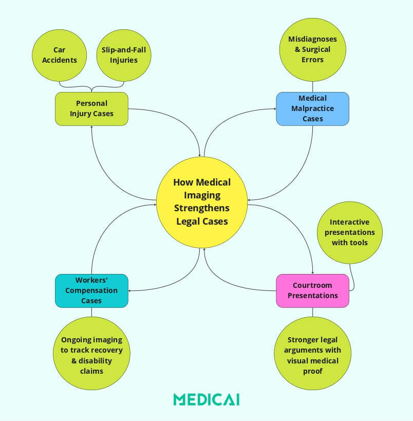
Motor vehicle accidents. Injuries frequently present without external signs. X-ray identifies fractures. MRI documents whiplash-associated soft tissue injury and disc herniation. CT identifies traumatic brain injury and internal bleeding in the acute phase. Emergency department imaging obtained hours after a collision is particularly valuable because its timing is documented and its purpose was clinical rather than forensic.
Premises liability and falls. CT confirms head trauma and intracranial bleeding. MRI documents spinal injury and disc damage. Ultrasound identifies internal bleeding that may not produce immediate symptoms. In fall cases, the causation question often turns on whether imaging findings are consistent with the mechanism of injury described, which is a question for the expert rather than the file.
Medical malpractice. Comparative imaging across time is the core evidence. A retained surgical instrument appears on post-operative imaging. A tumor visible on an earlier study but not reported supports a failure-to-diagnose claim. Serial imaging documenting progression while a provider took no action supports failure-to-treat. Malpractice cases typically require obtaining imaging from multiple providers across a treatment timeline, which makes retrieval logistics more demanding than in a single-incident case.
Workers’ compensation. Imaging tracks whether an injury has resolved, stabilized, or worsened, which drives disability determination. Serial studies document healing progress or the absence of it. Because workers’ compensation matters often continue for years, the firm’s ability to retrieve and organize imaging from across that period matters as much as the content of any single study.
Requesting medical images from providers
Retrieval is where most imaging problems originate, and most are avoidable.
Start with a valid authorization. A HIPAA-compliant authorization signed by the client is the foundation of the request. Authorizations that are incomplete, expired, or insufficiently specific are the most common reason providers reject or delay requests.
Request the images, not only the reports. A request for “medical records” frequently produces the radiology report and nothing else. The report is the radiologist’s interpretation; the images are the underlying evidence. Retaining experts to review the study requires the study itself. Specify that the request covers imaging files in DICOM format.
Be specific about modality, body part, and date range. “All imaging” invites either an incomplete response or a very slow one. Naming the modality (MRI, CT, X-ray), the anatomical region, and the date window produces faster and more complete production.
Request secure electronic delivery. Providers still default to burning studies onto CDs, which introduces delay, risk of loss, and the possibility that the disc is unreadable when it arrives. Where the provider supports secure electronic transfer, requesting it removes several failure points.
Track the request. Providers have statutory response deadlines, but practical compliance varies. A tracked request with documented follow-up supports a motion to compel where one becomes necessary.
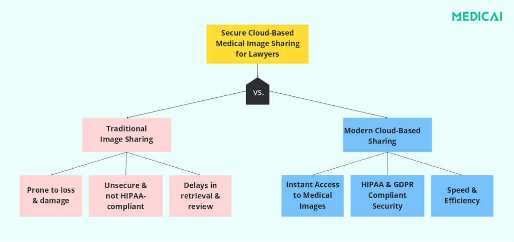
Image authentication and chain of custody
Imaging is subject to the same authentication requirements as any other documentary evidence. Opposing counsel can and does challenge whether a file is what it purports to be, whether it has been altered, and whether it corresponds to the plaintiff at all.
What DICOM metadata provides
Medical imaging is stored in DICOM format, which embeds structured metadata alongside the pixel data. That metadata typically records patient identifiers, study date and time, the acquiring institution, the specific device including manufacturer and model, acquisition parameters, and the referring physician.
This creates a contemporaneous record within the file itself. The study date is not a filename that can be changed; it is a data element written at acquisition. The device identifier ties the study to specific equipment at a specific facility. Where a study has been modified, DICOM implementations generally record that modification rather than silently overwriting.
For authentication purposes, this means the file carries internal evidence of its own provenance. An expert can testify to what the metadata shows and whether it is consistent with the clinical history.
What chain of custody requires
Chain of custody for imaging means being able to account for the file from the point of production by the provider to its presentation in court.
That requires knowing who received the study and when, where it has been stored, who has accessed it, whether any copy was modified, and who it was transmitted to outside the firm.
Traditional handling defeats this. A CD that circulates through an office, gets copied to a paralegal’s desktop, is emailed to an expert, and is later re-copied for opposing counsel produces no record of any of those events. If authenticity is challenged, the firm has testimony but no documentation.
Platforms that log access automatically resolve this by producing a timestamped record each time a study is viewed, downloaded, or shared, with the identity of the user. Where storage is tamper-evident, any modification attempt is recorded rather than silent. That record is what supports a foundation argument if authenticity is contested.
Practical handling rules
Do not rename files or alter file contents. Do not work from a copy when the original is available. Log transfers to experts and opposing counsel. Store studies in a single controlled location rather than distributed across individual machines. Retain the original production from the provider unmodified, and work from copies where working copies are needed.
HIPAA obligations for law firms
Law firms handling medical imaging take on obligations under HIPAA, and the mechanism by which this happens is often misunderstood.
HIPAA applies directly to covered entities: providers, health plans, and clearinghouses. It reaches other parties through the business associate relationship. A law firm that receives, stores, or transmits Protected Health Information on behalf of a covered entity, for example, defense counsel retained by a hospital, is generally a business associate and is directly subject to Security Rule requirements and to enforcement.
A plaintiff’s firm receiving records under client authorization is in a different position, since it acts on behalf of the individual rather than the covered entity. But this distinction does not eliminate exposure. State privacy statutes, professional responsibility rules governing confidentiality, and common law duties all apply to client health information regardless of HIPAA’s technical reach. Firms handling imaging should assume that the practical standard for handling PHI is the one HIPAA describes.
The safeguards that matter are consistent regardless of the analysis: encryption in transit and at rest, access controls restricting who in the firm can open a given study, audit logging of access, and written agreements with any vendor storing or transmitting the files.
For the full treatment of what HIPAA requires, including the Privacy Rule, Security Rule technical safeguards, business associate agreements, breach notification, and de-identification, see HIPAA-compliant medical image sharing.
Penalty exposure
HIPAA civil penalties are tiered by culpability, ranging from unknowing violations at the low end to uncorrected willful neglect at the high end, with per-violation minimums and maximums and an annual cap per violation category. HHS adjusts these figures for inflation annually, so current amounts should be verified against the HHS enforcement page rather than relied on from secondary sources.
For firms handling matters involving EU or EEA residents, GDPR applies independently, with penalties reaching the greater of €20 million or 4% of global annual turnover, and additional requirements for cross-border transfer including Standard Contractual Clauses or an adequacy decision.
Working with medical experts
Imaging becomes persuasive through expert interpretation, and the logistics of getting studies to experts affect both cost and case timeline.
Experts need the actual DICOM study rather than printed images or screenshots, because interpretation requires the ability to adjust window and level settings, scroll through slices, and measure. A printed image is a single fixed view of a study that contains hundreds.
Providing experts with browser-based access to the study avoids requiring them to install specialized software and avoids sending files that then exist outside the firm’s control. Where the platform logs access, the expert’s review is documented, which is occasionally useful when the extent of their review is challenged.
Retaining a radiologist rather than only a treating physician is worth considering where imaging interpretation is contested. Treating physicians testify to the patient’s course; a radiologist testifies to what the study shows and whether the original interpretation was sound.
Presenting imaging at deposition and trial
Imaging that persuades a jury is imaging they can understand.
Annotation. Marking the relevant finding directly on the image removes the ambiguity of an expert gesturing at a screen. Annotations should be clearly identified as such rather than appearing to be part of the original study.
Comparison views. Placing pre-incident and post-incident studies side by side, or serial studies in sequence, makes progression visible in a way that sequential description does not. This is frequently the most persuasive presentation in disc injury and malpractice matters.
Reconstruction. Three-dimensional reconstruction from CT data can make fracture patterns and internal injury comprehensible to a lay audience where axial slices are not. Where reconstruction is used, the expert should be able to explain how it was generated and confirm that it accurately represents the underlying data, since a reconstruction is a rendering rather than a photograph.
Preparation for challenge. Assume that any presentation of imaging will be met with argument about what the images show, whether the annotations are fair, and whether the presentation format overstates the finding. Presentations should be defensible on each point.
Firm policy and staff training
Most imaging mishandling is procedural rather than technical, and firm-level policy prevents more of it than any individual tool.
Designate responsibility. Naming a person accountable for imaging handling and privacy compliance, even in a small firm, produces a consistent point of decision rather than ad hoc handling.
Write the handling rules down. How imaging is requested, where it is stored, who may access it, how it is transmitted to experts and opposing counsel, and how it is retained or destroyed at matter close. Unwritten practice varies depending on who is doing it. Plenty of firms now draft that policy with an AI assistant, which quickly produces a complete first version but tends to return abstract subjects, formulaic transitions, and evenly paced sentences that staff skim rather than read. Passing the draft through a tool for humanizing AI text restructures those sentences rather than substituting synonyms, so the rules read like instructions written inside your own firm and are easier for a paralegal to follow under deadline pressure.
Train annually and on intake. Staff needs to know both the rules and the reason for them. Training that explains why unencrypted email is a problem produces better compliance than training that only prohibits it.
Prohibit the common workarounds explicitly. Personal email accounts, consumer file-sharing services, unencrypted USB drives, and personal cloud storage. These get used under deadline pressure unless the sanctioned alternative is faster.
Audit periodically. Reviewing access logs occasionally identifies both security problems and workflow problems worth fixing.
Technology for legal imaging workflows
Three categories of tools address the practical problems.
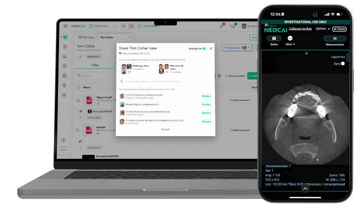
Cloud-based imaging platforms provide storage with access control and logging, remove the physical media problem, and allow controlled sharing with experts and opposing counsel through links rather than file transfers. Access logging is the feature that matters most for chain of custody.
Browser-based DICOM viewers allow attorneys, experts, and clients to review studies without installing software. Viewing rather than downloading also keeps the file within the platform’s audit trail. Some viewers include de-identification tools, which are relevant where studies are used in demonstrative exhibits or shared beyond the immediate case team.
Case management integration connects imaging to the matter file so studies are located through the same system as other case documents, which reduces the copies-scattered-across-desktops problem that defeats chain of custody.
Medicai provides cloud-based imaging with browser access, role-based permissions, and access logging. Other platforms serve this market as well, and firms should evaluate against their own volume, matter types, and existing case management stack.
Frequently Asked Questions on Medical Image Sharing for Lawyers
Can lawyers access medical images under HIPAA?
Yes, with valid patient authorization. A HIPAA-compliant authorization signed by the client permits the provider to release imaging to the firm. Defense counsel retained by a covered entity typically accesses PHI as a business associate under a written agreement instead. In either case the firm takes on obligations to safeguard the information after receipt.
How do lawyers request medical images from a hospital?
Submit a HIPAA-compliant authorization signed by the client, specifying the modality, body part, and date range, and stating explicitly that the request covers imaging files in DICOM format rather than reports alone. Request secure electronic delivery where available. Track the request against the provider’s statutory response deadline.
What is DICOM and why does it matter in litigation?
DICOM is the standard format for medical imaging, storing the images together with metadata recording the study date, acquiring facility, device, and acquisition parameters. That metadata supports authentication because it is written at acquisition rather than applied afterward, providing internal evidence of the file’s provenance if authenticity is challenged.
How do you establish chain of custody for medical images?
Document who received the study and when, where it is stored, every person who accessed it, whether any copy was modified, and every transmission outside the firm. Platforms that log access automatically produce this record. Files circulated on CDs and by email generally cannot support a documented chain.
Are medical images admissible as evidence?
Medical imaging is generally admissible subject to authentication and relevance, usually introduced through expert testimony. Challenges typically go to whether the file is what it purports to be, whether it has been altered, and whether any annotation or reconstruction fairly represents the original study.
Can a law firm store medical images in the cloud?
Yes, provided the platform offers appropriate safeguards including encryption, access controls, and audit logging, and provided there is a written agreement with the vendor covering its handling of the information. Consumer file-sharing services generally do not meet this standard on their default tiers.
What happens if a firm mishandles medical images?
Consequences may include HIPAA enforcement where the firm is a business associate, liability under state privacy statutes, professional responsibility exposure for breach of client confidentiality, and evidentiary consequences if mishandling undermines authentication. The evidentiary risk is frequently the most immediate: a study that cannot be authenticated may not reach the jury.
Do lawyers need special software to view medical images?
Not necessarily. DICOM files require a viewer rather than standard image software, but browser-based viewers allow review without local installation. This also keeps the study within the platform’s audit trail rather than creating copies on individual machines.
Conclusion
Medical imaging is frequently the strongest evidence in an injury matter, and the difference between imaging that carries weight and imaging that gets excluded usually comes down to handling rather than content. Requesting the study rather than only the report, preserving the original production unmodified, maintaining a documented record of access, and being able to explain the file’s provenance are what allow the evidence to do its work.
The handling requirements are not primarily technical. They are procedural, and firms that write down how imaging is handled and train staff on it have fewer problems than firms relying on individual judgment under deadline pressure.
For the underlying compliance framework, see HIPAA-compliant medical image sharing. For how medical image sharing works technically, see the medical image sharing guide.


Related Articles



Lets get in touch!
Learn more about how Medicai can help you strengthen your practice and improve your patients’ experience. Ready to start your Journey?
Book A Free Demo