PET-CT Scan: What It Is, Why It's Done, and What to Expect

A PET-CT scan combines positron emission tomography (PET) and computed tomography (CT) in a single machine, so doctors can see how tissues are functioning and exactly where they are in the body. It is most often used to find, stage, and monitor cancer, and also to study the heart and brain. The visit usually takes about two hours, of which the scan itself is about 30 minutes.
This guide explains how PET-CT works, why it is ordered, how to prepare, what happens on the day of the scan, radiation and side effects, and how to get your results and images.
What is a PET-CT scan?
A PET-CT scan is a single test that produces two kinds of images at once and overlays them. RadiologyInfo describes it this way: “Image fusion allows the doctor to connect and interpret information from two different exams on one image” (RadiologyInfo).
- PET shows activity. Before the scan, you receive a small amount of a radioactive tracer. “The most common radiotracer is F-18 fluorodeoxyglucose (FDG), a molecule similar to glucose” (RadiologyInfo). Cells that use a lot of sugar, including many cancer cells, take up more FDG and appear brighter on the PET image.
- CT shows anatomy. The computed tomography (CT) part uses X-rays to create detailed cross-sectional pictures of organs and bones.
Together, they show both what is happening and where. A bright spot on PET can be matched to a specific organ or lymph node on CT.
Why is a PET-CT scan done?
Doctors order a PET-CT scan to answer questions that anatomy alone cannot, such as whether a mass is active, whether cancer has spread, or whether treatment is working. The three main areas are cancer, heart disease, and brain disorders.
- Cancer. PET-CT is used to find cancer, see how far it has spread (staging), check whether treatment is working, and look for recurrence after treatment ends. The American Cancer Society lists uses including to “see if a current treatment is working” and “check for new cancer growth or recurrence after treatment ends” (ACS).
- Heart disease. PET can show blood flow to the heart muscle and help assess coronary artery disease and damage after a heart attack (Johns Hopkins Medicine).
- Brain disorders. PET helps evaluate brain tumors, epilepsy, dementia, and Alzheimer’s disease. It can help “find where seizures start in the brain” (Mayo Clinic).
What does a PET-CT scan show in cancer?
In cancer care, PET-CT shows areas where cells are taking up more of the tracer than surrounding tissue. Those areas may be tumors, lymph nodes, or other spots where cancer has spread. Cleveland Clinic lists breast, lung, and thyroid cancer among the cancers it is used for (Cleveland Clinic), and Johns Hopkins notes its role in lung cancer.
A bright area is not always cancer. Inflammation, infection, and recent surgery or radiation can also take up the tracer (see the limitations section below). That is why a specialist interprets the scan together with your history and other tests.
How to prepare for a PET-CT scan
Preparing for a PET-CT scan mostly means keeping your blood sugar steady, so the tracer goes where it should. Your imaging center will give you exact instructions. These are the typical ones:
- Fasting. Mayo Clinic advises not eating or drinking for about 4 to 6 hours before the scan (Mayo Clinic). Some centers ask for longer. Ask your center whether you may drink water.
- Exercise. Avoid heavy exercise for at least a day before the scan (Mayo Clinic).
- Diabetes. Ask for specific instructions, because your provider may change the fasting and medication plan (Cleveland Clinic). Some centers ask you not to take diabetes medication on the morning of the test (Stanford Health Care).
- Pregnancy and breastfeeding. Tell your provider if you are or might be pregnant, or if you are breastfeeding (Cleveland Clinic).
- Clothing and metal. Wear comfortable clothes and leave jewelry and other metal items at home (Cleveland Clinic).
- Allergies and medical history. Tell the team about any allergies, especially to CT contrast.
On the day of the test, your blood sugar is usually checked before the tracer is administered. “If your blood sugar is too high, you may need to reschedule your test” (ACS). Each center sets its own acceptable range. Memorial Sloan Kettering, for example, requires a level between 70 and 199 mg/dL (MSK).
What happens during a PET-CT scan
A PET-CT appointment follows the same five steps at most imaging centers.
- Check-in and blood sugar test. A technologist reviews your history and checks your blood sugar.
- Tracer injection. You receive the radioactive tracer, usually through an IV in your arm.
- Resting uptake period. You rest quietly while the tracer travels through your body. Cleveland Clinic describes this as sitting for about an hour, and Mayo Clinic says about 30 to 60 minutes (Mayo Clinic). Staying still and relaxed helps the tracer settle where it should.
- The scan. You lie on a table that moves slowly through a ring-shaped scanner. You need to lie still while the images are taken.
- After the scan. Most people can go home and return to normal activities. Drinking plenty of fluids helps flush the tracer out of your body faster.
How long does a PET-CT scan take?
The PET-CT scan itself takes about 30 minutes, and the whole visit takes about two hours. Most of that time is the resting uptake period before the scan. Some centers ask you to plan for 2 to 3 hours in total (Stanford Health Care). Scans that cover more of the body or include additional protocols can take longer.
PET-CT radiation dose, risks, and side effects
PET-CT is generally safe, and side effects are rare. It involves more radiation than many other imaging tests, which your doctor weighs against the value of the information it provides.
Radiation dose
A whole-body PET-CT exposes you to about 22.7 millisieverts (mSv) of radiation. That is roughly the amount of natural background radiation a person receives in 7.6 years (RadiologyInfo). For comparison, RadiologyInfo lists a chest X-ray at 0.1 mSv. The tracer “leaves your body within a few hours” (Cleveland Clinic).
Side effects and allergic reactions
Most people feel nothing unusual during or after a PET-CT. “Allergic reactions to radiotracers are extremely rare and usually mild” (RadiologyInfo). If your scan uses CT contrast dye, reactions to the contrast are also possible, such as rash, nausea, or an itchy throat (ACS). Tell the team right away if you feel unwell. You may also feel brief discomfort at the IV site.
Pregnancy, breastfeeding, and close contact
- Pregnancy. Tell your provider before the scan if you are pregnant or might be. Your doctor will decide whether the scan should go ahead or wait.
- Breastfeeding. “Radiation may pass through breast milk to a baby” (Mayo Clinic). RadiologyInfo suggests pumping milk ahead of time to use until the tracer and any contrast have left your body (RadiologyInfo). Some centers give a specific window. Memorial Sloan Kettering, for example, advises stopping breastfeeding for 4 hours after the scan (MSK). Follow your center’s instructions.
- Close contact. Some centers advise avoiding close contact with pregnant people and young children for a short time after the scan. MSK, for example, specifies 1 hour. Ask your care team what applies to you.
Benefits and limitations of PET-CT
PET-CT’s main benefit is that it shows how tissue is behaving and where it is, in one visit. Its main limits are that bright spots are not always cancer, and that image detail is lower than CT or MRI alone.
Benefits
- Function and anatomy together. Fused images allow doctors to match an active area on PET to its exact location on CT (RadiologyInfo).
- Whole-body view in one scan. Useful for staging cancer and checking for spread.
- Tracks change over time. Comparing scans before and after treatment shows whether activity has gone down.
- One appointment instead of two. Both scans happen on the same table during the same visit.
Limitations
- Not every bright spot is cancer. “Areas of inflammation or infection, or areas that have changed because of recent surgery or radiation” can also take up the tracer (ACS).
- Blood sugar matters. “Altered blood sugar or blood insulin levels may adversely affect the test results” (RadiologyInfo).
- Lower resolution than CT or MRI. “The image resolution of nuclear medicine images may not be as high as that of CT or MRI” (RadiologyInfo). Very small areas can be hard to assess.
- Radiation. The combined dose is higher than CT alone (see above).
PET-CT vs. PET, CT, and MRI
PET-CT, PET, CT, and MRI each answer different questions. Your doctor chooses based on what needs to be seen.
| Scan | What it mainly shows | Radiation | Common uses |
|---|---|---|---|
| PET-CT | Tissue activity plus detailed anatomy, fused | Yes. About 22.7 mSv for a whole-body scan | Cancer staging and response, some heart and brain studies |
| PET alone | Tissue activity, with less anatomic detail | Yes, from the radiotracer | Less common today. PET is usually performed alongside CT (Mayo Clinic). |
| CT | Detailed anatomy of organs, bones, and vessels | Yes. For example, about 7.7 mSv for the abdomen and pelvis | Injuries, infections, many cancers, surgical planning |
| MRI | Detailed soft tissue anatomy | No ionizing radiation | Brain, spine, joints, soft tissue tumors |
Doses are from RadiologyInfo. For a deeper comparison of the two most common scans, see MRI vs. CT. For an overview of other imaging modalities, see our guide to radiology modalities.
PET-CT results and viewing your images
A PET-CT scan is interpreted by a specialist, and the results are sent to the doctor who ordered it. “A radiologist or other doctor specially trained in nuclear medicine will interpret the images and send a report to your referring physician” (RadiologyInfo).
- When results arrive. Interpretation usually takes 24 to 48 hours. Mayo Clinic notes that a written report may take a few days, depending on the medical center (Mayo Clinic).
- What SUV means. PET-CT reports often include standardized uptake values (SUVs). MD Anderson describes an SUV as “a tape measure for radioactive activity in the body”: higher numbers mean brighter spots, which “may indicate a malignancy”. Inflammation and infection can also produce bright spots (MD Anderson). Your doctor interprets SUVs alongside the images and your history, so a single number is not a diagnosis.
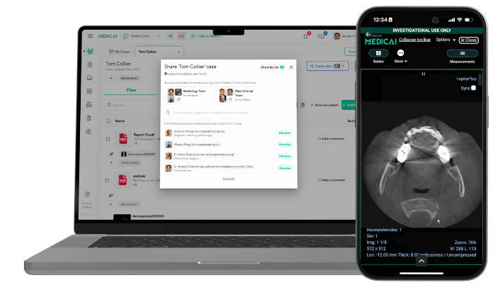
- Getting your images. Under HIPAA, you have a right to a copy of your images (HHS). They usually come as DICOM (.dcm) files, the standard format for medical images. You can open them in a browser with Medicai’s free online PET scan viewer, with no software to install.
- A second opinion. If you want another specialist to review your scan, see how a second opinion works.
Frequently Asked Questions
The PET-CT scan itself takes about 30 minutes, and the whole visit usually takes about two hours. Most of that time is the resting period after the tracer injection, which lasts about 30 to 60 minutes. Some centers ask patients to plan for 2 to 3 hours in total.
Before a PET-CT scan, do not eat for the time your center specifies, often about 4 to 6 hours, and avoid heavy exercise for at least a day. Do not skip telling your team if you have diabetes, are pregnant or breastfeeding, or have allergies to contrast. Leave jewelry and other metal items at home.
PET-CT is generally safe, and allergic reactions to the tracer are extremely rare and usually mild. It does involve radiation: a whole-body PET-CT is about 22.7 mSv, roughly 7.6 years of natural background radiation. Your doctor orders it when the information it provides outweighs that risk.
A PET-CT scan shows which tissues are taking up more of the radioactive tracer, matched to detailed anatomy from CT. In cancer care, this helps find tumors, see whether cancer has spread, and check whether treatment is working. Bright areas can also come from inflammation, infection, or recent surgery, so a specialist interprets the results.
A radiologist or nuclear medicine physician reads the scan and sends a report to the doctor who ordered it. Interpretation usually takes 24 to 48 hours, and a written report may take a few days depending on the medical center. Your doctor will explain the results with you.
Medicai helps patients and providers store, view, and share medical images, including PET-CT, through a cloud platform with a patient imaging portal and a free online PET scan viewer.

Related Articles


Lets get in touch!
Learn more about how Medicai can help you strengthen your practice and improve your patients’ experience. Ready to start your Journey?
Book A Free Demo