Radiology Modalities: From X-ray to Interventional

Every picture tells a story, but in medicine, the right picture can save a life. That’s the power of radiology, the science of seeing inside the body without a single incision.
Radiology modalities are the different imaging techniques doctors use to diagnose, monitor, and guide treatment. From X-rays and CT scans to MRI, ultrasound, and PET, each modality offers unique strengths, whether it’s spotting a fracture, mapping blood flow, or tracking a tumor.
Learn about the nine key radiology modalities, their uses, benefits, and limitations.

Radiology Modalities: An Overview
In radiology, a “modality” refers to a specific imaging technique used to view the body. Each modality, whether X-rays, sound waves, or magnetic fields, uses unique technology and provides different information to help doctors diagnose patients.
Radiology modalities can be grouped into two broad categories:
- Diagnostic imaging focuses on capturing images of the body to identify diseases, injuries, or abnormalities. Examples include X-rays, CT scans, MRI, ultrasound, and nuclear medicine.
- Interventional imaging extends beyond diagnosis by guiding minimally invasive procedures like biopsies, angioplasty, stent placements, and tumor ablations. This approach reduces pain, shortens recovery, and lowers surgical risks for patients.
Another important way to classify radiology modalities is by energy type:
- Ionizing modalities (X-ray, CT, PET, nuclear medicine) use radiation for imaging. While effective for many conditions, they necessitate careful dose management for patient safety.
- Non-ionizing modalities, like MRI and ultrasound, use magnetic fields, radio waves, or sound waves instead of radiation, making them preferable for children, pregnant patients, or cases needing repeated imaging.
No single imaging modality is superior. Each serves a unique purpose, like identifying fractures, monitoring tumors, guiding catheters, or assessing organ function.

The 9 Major Radiology Modalities
Let’s study the nine key radiology modalities and their applications, advantages, and limitations.
X-ray (Radiography)
X-ray imaging is the oldest and most common radiology technique. It uses ionizing radiation to produce a two-dimensional image of internal structures, where dense materials like bones appear white and softer tissues appear darker.
How the Test Is Done
During an X-ray, the patient is placed between the X-ray machine and a detector plate, either standing, sitting, or lying down. A protective lead apron may be used to shield sensitive areas from radiation. Patients must remain still for a few seconds to ensure clear images.
X-rays are used for all ages, but extra care is taken with children and pregnant women due to radiation exposure.
Common Uses
- Detecting bone fractures and dislocations
- Identifying chest infections like pneumonia
- Assessing arthritis and joint changes
- Evaluating dental issues (cavities, impacted teeth)
- Spotting fluid buildup or foreign objects in the body
Duration
- Exposure lasts a fraction of a second; the full process takes 5–15 minutes.
Advantages
- Fast and efficient – results within minutes
- Inexpensive compared to other methods
- Widely available almost everywhere
Limitations
- Limited soft-tissue detail
- Radiation exposure, though minimal
Fluoroscopy
Fluoroscopy uses a continuous X-ray beam to create real-time images of internal structures in motion, unlike a standard X-ray that provides just a single snapshot. This technique is especially useful for guiding procedures and assessing organ function during activity.
How the Test Is Done
The patient lies on a fluoroscopy table as an X-ray source projects radiation to a monitor. They may need to swallow a contrast material, such as barium, or receive a contrast dye injection. The radiologist monitors the patient’s movements and may request adjustments in position.
Fluoroscopy is suitable for all ages, but radiation doses are carefully controlled for children and pregnant women.
Duration
Diagnostic studies typically take 15–30 minutes, while interventional procedures may last 1–2 hours, depending on complexity.
Common Uses
- Barium swallow or GI tract exams (esophagus, stomach, intestines)
- Cardiac catheterization and stent placement
- Guiding needles, catheters, or biopsies in real time
- Checking joint movement or orthopedic alignment
Advantages
- Provides dynamic, real-time imaging
- Essential for interventional radiology procedures
- Allows visualization of both structure and function
Limitations
- Higher radiation exposure compared to a single X-ray
- Requires contrast materials, which may pose allergy risks
- Needs specialized equipment and trained staff
Computed Tomography (CT)
Computed Tomography (CT) uses X-rays and advanced computer processing to create detailed cross-sectional images of the body. CT produces slices that can be reconstructed into 3D images, making it a versatile imaging tool in modern medicine.
How the Test Is Done
The patient lies on a motorized table that moves through a circular CT scanner, where a rotating X-ray tube captures images from various angles to create cross-sectional slices. A contrast dye may be used to enhance the visibility of organs, blood vessels, or tumors.
Patients must stay still and may need to hold their breath briefly during the scan.
CT is suitable for all ages, but risks are considered for children and pregnant women due to radiation.
Duration
A standard CT scan usually takes 5–10 minutes, while specialized scans with contrast may take up to 30 minutes, including preparation.
Common Uses
- Detecting internal bleeding and trauma injuries
- Identifying and staging tumors and cancers
- Diagnosing lung diseases such as pneumonia or pulmonary embolism
- Evaluating blood vessels for aneurysms, blockages, or clots
- Planning surgeries or radiation therapy
Advantages
- Provides detailed images of bone, soft tissue, and blood vessels
- Fast and reliable – often used in emergencies
- Can produce 3D reconstructions for surgical planning
Limitations
- Higher radiation dose compared to standard X-rays
- Contrast dye risks – possible allergic reactions or kidney complications
- Less effective than MRI for soft-tissue detail in the brain and spinal cord
Magnetic Resonance Imaging (MRI)
Magnetic Resonance Imaging (MRI) uses strong magnets and radio waves to create detailed images of soft tissues without radiation. It’s especially useful for repeated scans of the brain, spine, joints, and internal organs, and can detect conditions often missed by other imaging methods.

How the Test Is Done
During an MRI, the patient lies on a table that slides into a tunnel-like scanner. The machine uses magnetic fields and radiofrequency pulses to produce detailed images.
Sometimes, a gadolinium-based contrast agent is injected to enhance the visibility of blood vessels, tumors, or inflammation.
Patients must remove all metal objects (jewelry, watches, piercings, hairpins) during the test.
Duration
Most MRI exams take 30–60 minutes, while complex studies may last up to 90 minutes.
Common Uses
- Diagnosing brain tumors, strokes, and multiple sclerosis
- Evaluating spinal cord injuries and disc problems
- Detecting soft tissue injuries in muscles, ligaments, and tendons
- Cardiac MRI for heart structure and function
- Assessing abdominal and pelvic organs (liver, uterus, prostate, etc.)
Advantages
- No radiation exposure
- Provides superior soft-tissue detail compared to CT
- Wide variety of specialized applications (fMRI, MRA, spectroscopy)
Limitations
- Expensive and less available than X-ray or CT
- Longer scan times, requiring patient cooperation
- Not suitable for patients with implants or severe claustrophobia
Ultrasound (Sonography)
Ultrasound imaging uses high-frequency sound waves to create real-time images of internal organs and tissues without radiation. It is a safer option for pregnant women and children.
It works by sending sound waves through the body, which bounce back from tissues or fluids. A computer then converts the echoes into images.
How the Test Is Done
During an ultrasound, the patient lies on a bed with a clear gel applied to the skin. A handheld device called a transducer is moved over the area, sending sound waves into the body and capturing the echoes.
For certain ultrasounds, like transvaginal or transesophageal scans, a specialized probe may be inserted for better imaging.
Duration
Most ultrasound exams last 15–30 minutes, depending on the type of study.
Common Uses
- Monitoring pregnancy and fetal growth
- Abdominal imaging (liver, gallbladder, kidneys, pancreas)
- Detecting blood clots and evaluating blood flow (Doppler)
- Echocardiography for heart conditions
- Guiding biopsies, drainages, or injections
Advantages
- No radiation exposure
- Safe and non-invasive for all ages
- Portable and widely available
- Provides real-time imaging
Limitations
- Operator-dependent – image quality depends on skill
- Limited in patients with obesity or excess gas
- Cannot penetrate bone or air-filled organs (like lungs)
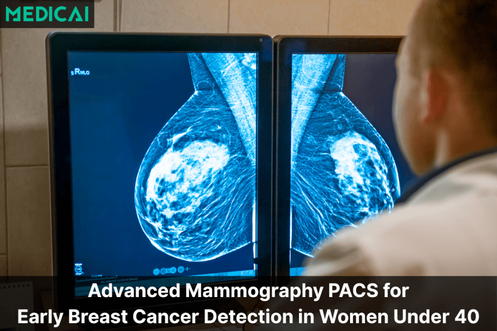
Mammography
Mammography is a low-dose X-ray technique crucial for early breast cancer detection, revealing abnormalities before physical exams. Modern digital and 3D mammography offer clearer, detailed images with lower radiation exposure.
How the Test Is Done
During a mammogram, the patient stands in front of the unit, placing each breast on a flat plate. A second plate compresses the breast to improve image clarity and reduce radiation dose. Images are captured from various angles, with brief compression lasting a few seconds per image.
Mammography is recommended for women aged 40 and older, with earlier screening for those at high risk.
Duration
Usually takes 15–20 minutes.
Common Uses
- Routine breast cancer screening in eligible women
- Evaluating lumps, pain, or nipple discharge
- Monitoring women with a history of breast cancer
- Guiding breast biopsies for suspicious findings
Advantages
- Proven tool for early breast cancer detection
- Detects microcalcifications that may indicate early disease
- Widely available in most healthcare settings
Limitations
- Radiation exposure, though minimal
- Less effective in dense breast tissue
- May cause temporary discomfort from compression
Nuclear Medicine (Scintigraphy & SPECT)
Nuclear medicine is an imaging technique that emphasizes organ function over structure. It uses small amounts of radioactive materials, or radiotracers, which emit gamma rays that are captured by cameras to create functional images of organs.
SPECT (Single Photon Emission Computed Tomography) enhances this by providing 3D imaging for more detailed analysis.
How the Test Is Done
Radiotracers vary by organ; bone scans typically use injected tracers, while thyroid scans may involve swallowing a capsule. After the tracer accumulates, the patient lies still on a table while a gamma camera or SPECT scanner captures images.
The scan is painless, but stillness is essential for clear results.
Nuclear medicine is used in both adults and children, with care taken to minimize radiation exposure.
Duration
The scan usually lasts 30–60 minutes, depending on the tracer and test type, with some studies requiring delayed imaging later.
Common Uses
- Bone scans for fractures, infections, or metastatic cancer
- Thyroid imaging for hyperthyroidism or nodules
- Cardiac perfusion scans to check blood flow to the heart
- Kidney scans for function and obstruction evaluation
Advantages
- Provides functional information that other imaging cannot
- Detects disease early, often before structural changes appear
- Useful for whole-body imaging
Limitations
- Involves radiation exposure from tracers
- Lower resolution compared to CT or MRI
- Limited availability of certain specialized tracers
Positron Emission Tomography (PET)
Positron Emission Tomography (PET) is a nuclear imaging technique that shows metabolic and cellular activity in tissues, emphasizing organ and cell function over structure. It is particularly effective for detecting cancer, assessing brain disorders, and evaluating heart health.
How the Test Is Done
A small amount of a radioactive tracer, usually FDG (fluorodeoxyglucose), is injected into a vein. The tracer collects in areas with high metabolic activity, like rapidly growing tumors.
After 30–60 minutes, the patient lies on a scanning table while the PET scanner records signals to create detailed functional images.
Duration
Patients should expect the entire process to take 1–2 hours, including 30–60 minutes for preparation and uptake, followed by a 20–30 minute scan.
Common Uses
- Cancer detection, staging, and monitoring treatment response
- Neurological imaging for Alzheimer’s, Parkinson’s, and seizure focus mapping
- Cardiac evaluation for blood flow and damaged heart tissue
- Identifying inflammation or infection in certain conditions
Advantages
- Extremely sensitive for detecting disease activity
- Provides functional and metabolic information not seen on CT or MRI
- Useful for monitoring treatment response over time
Limitations
- High cost and limited availability in some regions
- Requires a radiopharmacy for tracer preparation
- Provides lower anatomical detail (often paired with CT or MRI for precision)
Interventional Radiology (IR)
Interventional Radiology (IR) is a subspecialty that uses imaging guidance to perform minimally invasive procedures. This technique helps doctors to treat conditions with small incisions, using catheters, wires, and needles.
How the Procedure Is Done
Patients undergoing an IR procedure typically receive local anesthesia or mild sedation. The radiologist makes a small incision to insert a catheter or needle, using real-time imaging for precise targeting.
Contrast dye may be injected to visualize blood vessels or guide instruments.
Duration
Most IR procedures last 30 minutes to 2 hours, with some needing an overnight hospital stay for monitoring.
Common Uses
- Vascular procedures such as angioplasty or embolization
- Cancer treatments like tumor ablation or chemoembolization
- Image-guided biopsies of lungs, liver, or kidneys
- Drainage of abscesses or fluid collections
- Stopping internal bleeding in emergencies
Advantages
- Minimally invasive, requiring only small incisions
- Shorter recovery times compared to surgery
- Often performed under local anesthesia
- Reduces overall hospital stay and complications
Limitations
- Requires specialized expertise and equipment
- Small risk of bleeding, infection, or injury at the site
- May not be suitable for all conditions
Conclusion
Radiology modalities give doctors the power to see more, treat earlier, and plan better. From X-rays to MRI and PET, each modality offers unique insights that shape patient care. No single scan answers every question, but together, they build a clearer story.
That’s where platforms like Medicai make a difference by connecting, organizing, and streamlining these imaging tools in one place. With smarter access to imaging, healthcare teams can deliver faster, safer, and more accurate care.


Related Articles


Lets get in touch!
Learn more about how Medicai can help you strengthen your practice and improve your patients’ experience. Ready to start your Journey?
Book A Free Demo