AI in Neurology Imaging: Clinical Uses, FDA-Cleared Tools, and Limits

AI in neurological imaging is software, mostly based on deep learning, that detects, measures, or prioritizes findings on brain and spine CT and MRI scans. Today it is used in four main ways:
- Flagging suspected strokes and brain bleeds for faster review
- Measuring brain volumes and lesions in dementia and multiple sclerosis
- Outlining brain tumors
- Making MRI scans faster and clearer
Most of these tools are FDA-cleared as aids for trained clinicians. None of them replaces the neurologist or radiologist who makes the diagnosis.
This guide covers each use case and the FDA-cleared tools behind it, how AI results reach clinicians in the imaging workflow, and the limits that still apply. For AI across all of radiology, see our overview of AI in radiology.

How AI is used in neurology imaging
AI in neurology imaging is used in nine main clinical tasks, each backed by at least one FDA-cleared tool. The table lists examples, not every product on the market. Descriptions follow each device’s cleared indication, not vendor marketing.
| Clinical use | What the AI does | Modality | Example FDA-cleared tools (record, year) | Where it fits |
|---|---|---|---|---|
| Large vessel occlusion (LVO) | Flags a suspected LVO and notifies the stroke specialist | CT angiography | Viz LVO (K223042, 2022); Rapid LVO (K221248, 2022); Aidoc BriefCase (K192383, 2019) | Emergency triage, in parallel with the standard read |
| Intracranial hemorrhage (ICH) | Flags a suspected bleed so the study is read sooner | Non-contrast head CT | Aidoc BriefCase (K180647, 2018); Rapid ICH (K221456, 2022) | Worklist prioritization |
| ASPECTS scoring | Helps assess early ischemic change in the brain tissue | Non-contrast head CT | Brainomix e-ASPECTS (K221564, 2023); Rapid ASPECTS (K232156, 2024) | Stroke workup decision support |
| Perfusion analysis | Helps select patients with a known ICA or M1 occlusion for thrombectomy | CT or MR perfusion | RAPID (K182130, 2018) | Stroke team treatment decision |
| Brain volumetrics | Labels and measures brain structures and compares them with reference data | MRI (icobrain also covers non-contrast CT) | NeuroQuant (K241098, 2024); icobrain (K192130, 2019); cNeuro cMRI (K171328, 2018) | Dementia and MS evaluation, attached to the report |
| ARIA detection | Acts as a concurrent reading aid for amyloid-related imaging abnormalities | Brain MRI | icobrain aria (K240712, 2024) | Safety monitoring during anti-amyloid therapy |
| Brain tumor segmentation | Semi-automatically outlines and measures high-grade glioma | MRI | Neosoma NS-HGlio (K221738, 2022) | Planning and response tracking, not primary diagnosis |
| MRI enhancement | Reduces image noise or increases sharpness | MRI | SubtleMR (K191688, 2019); GE SIGNA Premier with AIR Recon DL (K193282, 2020) | During or right after acquisition |
| White matter tract mapping | Turns diffusion MRI into 3D tract maps and measurements, without diagnosing | Diffusion MRI | Imeka ANDI (K230913, 2023) | Quantitative review |
Records were checked on Sep 30, 2026. Several vendors have newer clearances for later versions. For the full, periodically updated list, see FDA’s Artificial Intelligence-Enabled Medical Devices page. For AI tools outside neurology, see our list of AI diagnosis vendors.
AI for stroke detection and triage
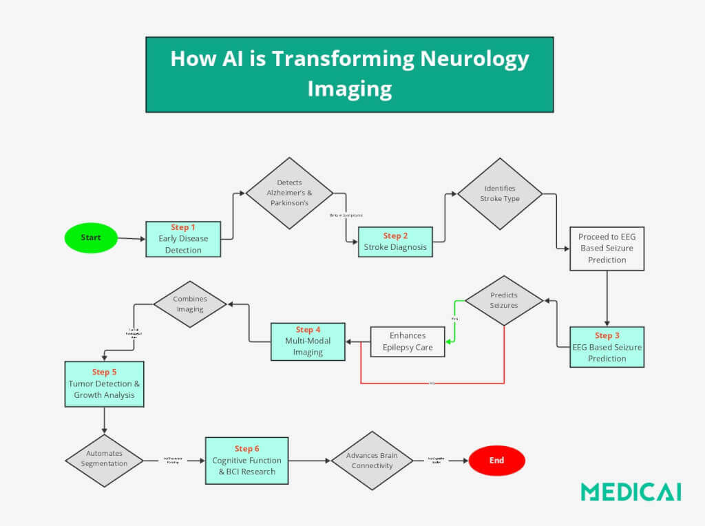
Stroke is where AI in neurology imaging is most established, because minutes matter. Stroke AI does not diagnose. It flags suspected findings so the right specialist can review the right scan sooner, and it supports the stroke team’s treatment decisions.
Large vessel occlusion detection and care team alerts
LVO detection software reads CT angiography of the head and alerts a neurovascular specialist when it suspects a blockage in a large artery, such as the internal carotid or the first segment of the middle cerebral artery (M1). The first tool of this kind, Viz.ai’s ContaCT, received FDA De Novo authorization in February 2018 (DEN170073). FDA describes it as a “notification-only, parallel workflow tool”: it sends an alert alongside the standard read and does not replace it. Viz.ai, RapidAI, and Aidoc now all have cleared LVO tools.
Intracranial hemorrhage detection
Hemorrhage detection software analyzes non-contrast head CT and flags suspected bleeding, so the study moves up the radiologist’s worklist. Rapid ICH, for example, is cleared to flag intraparenchymal, intraventricular, subarachnoid, and subdural hemorrhage. The radiologist still reads every study and makes the diagnosis. The AI changes the reading order, not the reader.
ASPECTS scoring and CT perfusion
ASPECTS (the Alberta Stroke Program Early CT Score) grades early ischemic change across 10 regions of the middle cerebral artery territory. Scoring by eye varies between readers. Brainomix e-ASPECTS and Rapid ASPECTS are cleared as computer-aided diagnosis tools that help clinicians assess these changes on non-contrast CT.
Perfusion software such as RAPID maps blood flow on CT or MR perfusion. It is cleared to help physicians select patients with a known ICA or proximal MCA occlusion for endovascular thrombectomy. The decision stays with the stroke team. For background on the two main scan types, see CT vs. MRI.
AI for dementia, multiple sclerosis, and neurodegeneration
In dementia and multiple sclerosis, AI’s main cleared role is measurement. It turns what radiologists judge by eye, such as how much a brain region has shrunk or how many lesions are new, into numbers that can be compared over time.
Brain volumetrics (quantitative MRI)
Volumetric software automatically labels brain structures on MRI, measures their volumes, and compares them with age-matched reference data. NeuroQuant, icobrain, and cNeuro cMRI are all FDA-cleared for this. Reports show, for example, whether the hippocampus is smaller than expected for the patient’s age, which supports a neurologist’s dementia workup. Volumetrics does not diagnose Alzheimer’s disease on its own. Amyloid and tau, the protein markers of Alzheimer’s, are measured with PET imaging or fluid tests, not routine MRI.
Multiple sclerosis lesion tracking
In multiple sclerosis, disease activity is tracked by counting and measuring white matter lesions across serial MRIs. Comparing a new scan with several priors by eye is slow, and small changes are easy to miss. Quantitative tools measure brain structure and lesions consistently from one scan to the next. That is why the same archive needs to keep every prior study available.
ARIA monitoring for anti-amyloid therapies
Anti-amyloid Alzheimer’s drugs have made MRI safety monitoring routine. They can cause amyloid-related imaging abnormalities (ARIA), which include brain swelling and small bleeds.
- The FDA label for Leqembi (lecanemab) calls for a baseline brain MRI and MRIs before the 3rd, 5th, 7th, and 14th infusions (FDA).
- The label for Kisunla (donanemab) calls for MRIs before the 2nd, 3rd, 4th, and 7th infusions (label).
icobrain aria is FDA-cleared as a concurrent reading aid that helps radiologists detect and characterize ARIA on these scans.
Research models that predict who will develop a neurodegenerative disease are not cleared for clinical use. For seizures and epilepsy, EEG remains the core test. See what an EEG shows that an MRI cannot.
AI for brain tumors
For brain tumors, AI’s cleared role is segmentation: outlining the tumor and measuring its volume on MRI. Manual outlining is slow, and measurements can vary between readers. Neosoma NS-HGlio, for example, is FDA-cleared for semi-automatic labeling and volume measurement of high-grade gliomas. Its indication states that it is not for primary diagnosis. Consistent tumor volumes help with surgical and radiation planning and with tracking response to treatment across follow-up scans. For how these models work, see our guide to deep learning MRI segmentation.
Radiomics, which extracts numerous quantitative features from images to predict tumor type, growth, or treatment response, is an active area of research. It is not yet a cleared clinical tool.
AI for faster, cleaner brain MRI
Deep learning also works before anyone reads the scan, by improving the images themselves. Brain MRI trades off scan time against image quality: longer scans give cleaner images, but patients struggle to stay still. Deep learning reconstruction and enhancement tools reduce noise or sharpen images, helping sites shorten scan times or obtain clearer images in the same scan time.
- Software added to existing scanners: SubtleMR is FDA-cleared to reduce image noise in head, spine, neck, and knee MRI, and to increase sharpness in non-contrast head MRI.
- Built into the scanner: GE HealthCare’s SIGNA Premier clearance includes AIR Recon DL, a deep learning reconstruction designed to improve signal-to-noise ratio and sharpness. Other scanner makers also market deep-learning reconstruction, so check each product’s clearance.
Whether an enhanced scan is appropriate for a given clinical question remains the radiologist’s call, and protocols should be validated locally. For contrast-enhanced studies, see brain MRI with contrast.
How AI fits into the neuroimaging workflow
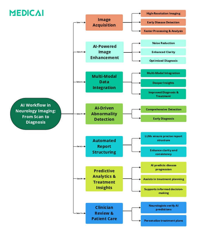
AI in neuroimaging is only useful if its output reaches the right clinician at the right moment, inside the tools they already use. Most neuro AI tools work in one of three ways:
- Triage and notification. Stroke and hemorrhage tools analyze a study as soon as it arrives and send alerts or reorder the worklist. The study still goes to the radiologist.
- Results inside the viewer. Volumetric reports, ARIA findings, and tumor outlines come back as additional DICOM series or reports stored with the original study. Clinicians view them alongside the images and compare them with priors.
- Reporting support. AI drafts or structures parts of the report for the radiologist to review and sign. See AI-assisted structured reporting.
All three depend on the PACS: how studies are routed to the AI, how results are returned, and whether neurologists outside the hospital can view them. Our guide to integrating AI into PACS covers the technical requirements.
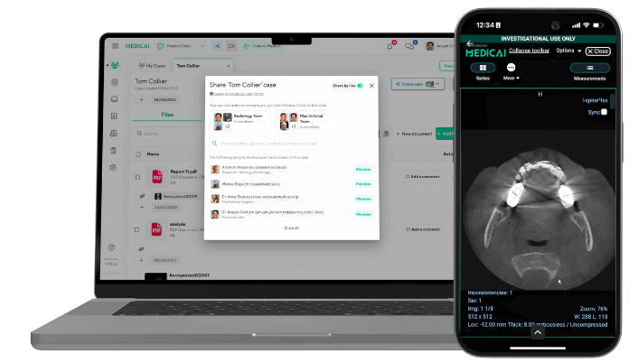
Medicai’s cloud PACS supports this workflow. Its Radiology AI Co-Pilot automates image sorting, preliminary analysis, and report generation. It also flags critical findings to prioritize studies that need immediate attention and converts dictations into structured notes. Neurology clinics such as NeuroAxis, a specialized neurology practice, use Medicai to manage their imaging.
Limits and risks of AI in neuroimaging
AI in neuroimaging has real limits: it depends on sensitive data, it can be hard to explain, it can perform unevenly across patient groups, and it changes faster than traditional devices. Buyers should ask vendors about each one.
Data privacy and security
Neuroimaging AI is trained and run on large sets of patient images that count as protected health information. In the US, that brings HIPAA obligations. In Europe, it brings GDPR obligations. Ask where images are processed, whether they leave your cloud or network, whether they are de-identified for training, and what the vendor’s business associate agreement covers.
The black box problem
Many deep learning models do not explain why they flagged a finding, which makes clinicians hesitant to trust them. Cleared triage tools address this partly by design: they alert a specialist, who still reviews the images. Explainable AI methods, such as heat maps showing which regions drove a result, are an active area of research. So are models that combine MRI, PET, and EEG to study brain connectivity in conditions such as depression and schizophrenia. Neither is standard clinical practice yet.
Bias across patient populations
A model trained mostly on one population can perform worse on others, such as different ages, ethnicities, sexes, scanners, or hospitals. Ask vendors what data their FDA submission used, which sites and scanners it included, and whether performance was reported by subgroup. Validate on your own patients before relying on a tool.
Regulation of changing AI models
Most imaging AI reaches the US market through FDA 510(k) clearance or De Novo authorization, not premarket approval (PMA). Models that keep learning used to fit poorly with rules written for fixed devices. In December 2024, the FDA finalized guidance on predetermined change control plans (PCCPs), which allows manufacturers to describe planned model updates in advance. FDA reissued the guidance in August 2025. In Europe, AI tools are regulated as medical devices under the EU MDR.
Frequently Asked Questions
In neurology imaging, AI is used mainly to flag suspected strokes and brain bleeds on CT for faster review, measure brain volumes and lesions on MRI for dementia and multiple sclerosis, help detect ARIA during anti-amyloid therapy, outline brain tumors, and improve MRI image quality. Most tools are FDA-cleared as aids for trained clinicians, who still make the diagnosis.
No. FDA-cleared neuroimaging AI tools are designed to assist trained clinicians, not replace them. Stroke tools send alerts or reorder worklists while the specialist still reads the scan, and measurement tools produce numbers that a physician interprets in clinical context. AI changes how quickly and consistently some tasks are done, not who is responsible for the diagnosis.
Most AI for brain imaging is FDA-cleared through the 510(k) pathway or authorized through De Novo, rather than approved through premarket approval (PMA). Examples include stroke triage tools from Viz.ai, RapidAI, and Aidoc, and brain volumetric tools such as NeuroQuant and icobrain. FDA keeps a periodically updated list of AI-enabled medical devices on its website.
Yes, as a triage aid. FDA-cleared tools analyze CT angiography for suspected large vessel occlusions and non-contrast head CT for suspected intracranial hemorrhage, then alert the care team or move the study up the worklist. A radiologist or stroke specialist still reviews the images and confirms the diagnosis.
Not on its own. FDA-cleared volumetric tools measure brain structures such as the hippocampus on MRI and compare them with age-matched reference data, which supports a neurologist’s dementia evaluation. Alzheimer’s diagnosis also relies on clinical assessment and on amyloid and tau biomarkers measured with PET imaging or fluid tests.
Medicai is a cloud PACS with a Radiology AI Co-Pilot, so neurology and radiology teams can store brain imaging, receive AI results alongside the original studies, and share them with clinicians wherever they read. To see how it fits your neuroimaging workflow, explore Medicai Cloud PACS or book a demo.



Related Articles
Lets get in touch!
Learn more about how Medicai can help you strengthen your practice and improve your patients’ experience. Ready to start your Journey?
Book A Free Demo