Medical Imaging Technology
The Medical Imaging Technology category highlights the latest advancements and innovations in medical imaging. This section covers topics such as the development of new imaging techniques, improvements in diagnostic accuracy, and the integration of technologies like artificial intelligence and cloud-based solutions. Articles explore how these innovations are transforming the way healthcare professionals capture, analyze, and utilize imaging data to improve patient care and streamline medical workflows.
136 posts
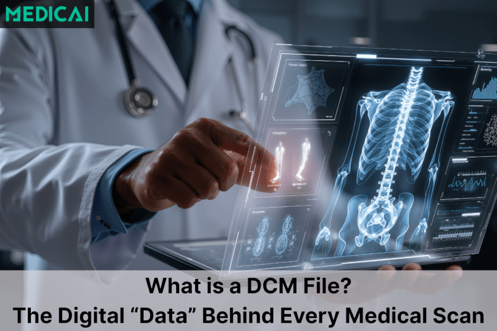
Read MoreDCM Files Explained: The Digital “Folder” Behind Every Medical Scan
Read MoreVNA in Radiology: How Vendor-Neutral Archives Fit Into the Radiology Workflow
Read MoreRadiology Modalities: From X-ray to Interventional

Read MorePACS System Radiology: Workflow, Benefits, and Challenges
Read MoreDICOM Web Viewer: How Zero-Footprint Browser-Based Viewing Works

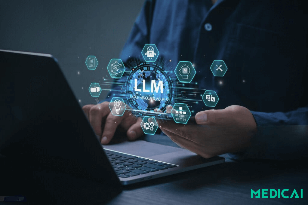
Read MoreHow Large Language Models Transform Radiology Today
Read More5 Best Free Mobile DICOM Viewers: Usability, Options, and Ways to pick the right one
Read MoreAI Radiology Workflow: How AI Fits Into Every Stage of Reading, Reporting, and Worklist Management
Read MoreHow Pediatric Teleradiology Transforms Children’s Healthcare

Read MoreTeleradiology PACS for Urgent Care Clinics — Cloud Imaging, Remote Reads, and 24/7 Coverage
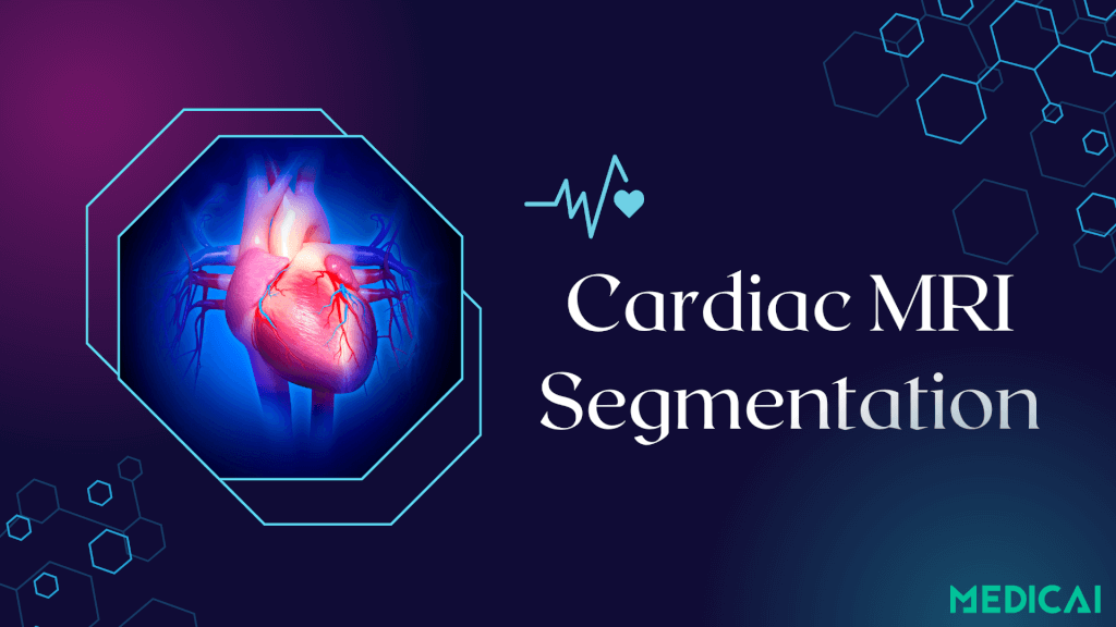
Read MoreFrom Manual to AI: The Future of Cardiac MRI Segmentation
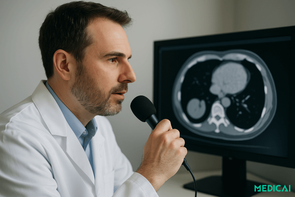
Read MoreVoice‑Enabled Radiology: From Dictation to Contextual Command