Page 4 of 22 – Recent Articles
449 posts
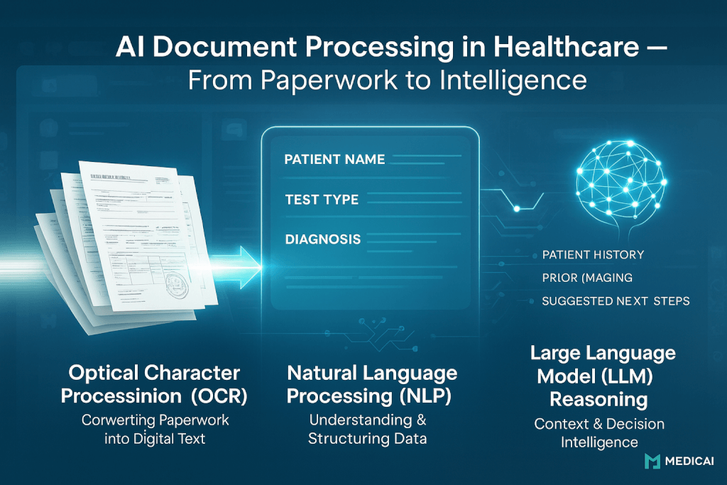
Bringing Order to Chaos: The Role of AI in Medical Document Processing
How AI Document Processing Is Transforming Healthcare Administration
HIPAA & GDPR Guide to Secure Medical Image Sharing

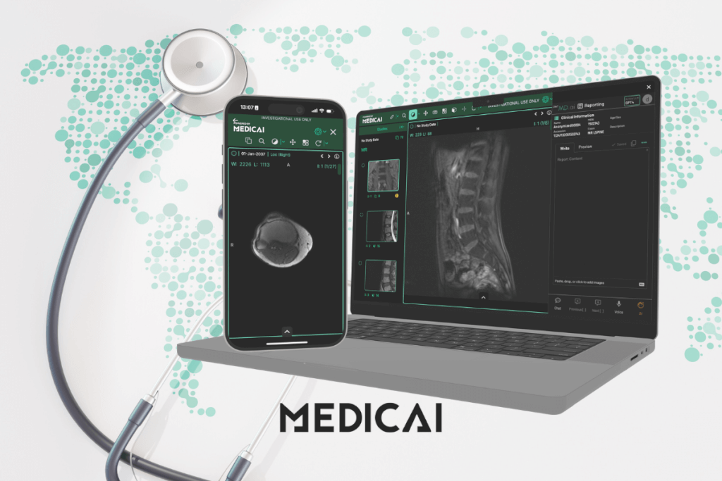
Simplify Imaging: The Power of Online DICOM Viewers
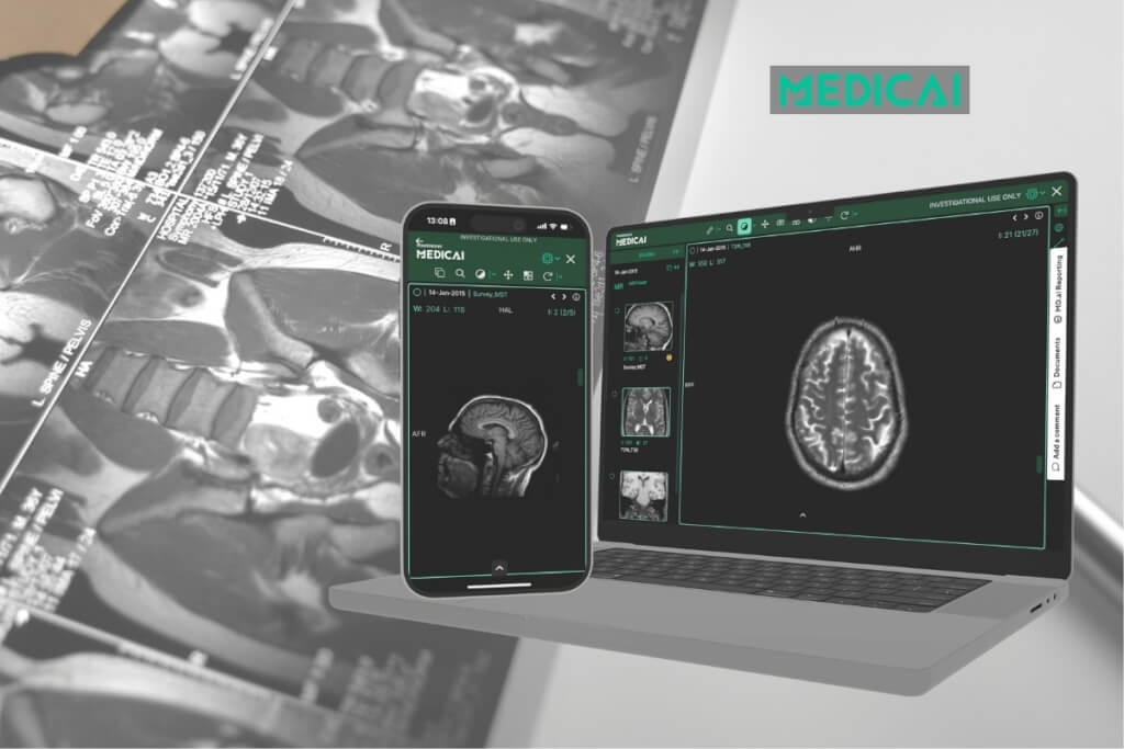
DICOM Viewer Features That Make Scans Simple

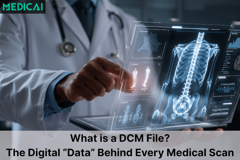
DCM Files Explained: The Digital “Folder” Behind Every Medical Scan
Specialty PACS Market: Trends, Growth, and Key Insights

VNA in Radiology: How Vendor-Neutral Archives Fit Into the Radiology Workflow

Radiology Modalities: From X-ray to Interventional

PACS System Radiology: Workflow, Benefits, and Challenges

DICOM Web Viewer: Smarter Imaging Anywhere

How Large Language Models Transform Radiology Today

5 Best Free Mobile DICOM Viewers: Usability, Options, and Ways to pick the right one
AI Radiology Workflow: How AI Fits Into Every Stage of Reading, Reporting, and Worklist Management
How Pediatric Teleradiology Transforms Children’s Healthcare

Teleradiology PACS for Urgent Care Clinics — Cloud Imaging, Remote Reads, and 24/7 Coverage

From Manual to AI: The Future of Cardiac MRI Segmentation

Voice‑Enabled Radiology: From Dictation to Contextual Command

Why Structured Annotations Are the Future of Oncologic Reporting?

How to Implement PACS in Urgent Care — Configuration, EHR Integration, and Best Practices

PACS Integration for Modern Veterinary Practices