Orthopedic Imaging: Modalities, Clinical Uses, and In-Office Workflow

Orthopedic imaging is the use of medical imaging to diagnose bone, joint, and soft-tissue conditions, plan orthopedic surgery, and assess how patients heal afterward. Five modalities do most of the work: X-ray, MRI, CT, musculoskeletal ultrasound, and DEXA. Fluoroscopy and nuclear medicine play supporting roles. Orthopedic surgeons, sports medicine physicians, and primary care clinicians order these studies. Radiologists or the treating physicians interpret them.
This guide compares the modalities, explains which ones are used for each body region, and walks through the imaging workflow from order to post-operative follow-up. It also covers what orthopedic practices need to bring imaging in-house.
What is Orthopedic Imaging?
Orthopedic imaging is diagnostic imaging of the musculoskeletal system (bones, joints, cartilage, ligaments, tendons, muscles, and the spine), ordered to answer a specific clinical question. Typical questions: Is the bone broken? Is the ligament torn? Is the implant in the right position? Orthopedic imaging differs from general radiology in two ways:
- It is task-directed. Most studies confirm or rule out a suspected diagnosis that the clinician has already formed based on the history and exam.
- It is workflow-integrated. The same images are used for diagnosis, surgical planning, and years of post-operative comparison.
Modality choice follows evidence-based guidance such as the ACR Appropriateness Criteria. These rate imaging options for scenarios like acute knee trauma and chronic shoulder pain.
Orthopedic imaging vs. musculoskeletal (MSK) imaging
Orthopedic imaging and musculoskeletal imaging cover the same anatomy from different sides. “Orthopedic imaging” is the term orthopedic surgeons, sports medicine physicians, and primary care clinicians use for the studies they order. “Musculoskeletal” or “MSK imaging” is the name of the radiology subspecialty. MSK radiologists complete fellowship training focused on interpreting these studies.
Imaging Modalities Used in Orthopedics: Five Primary and Two Adjunct
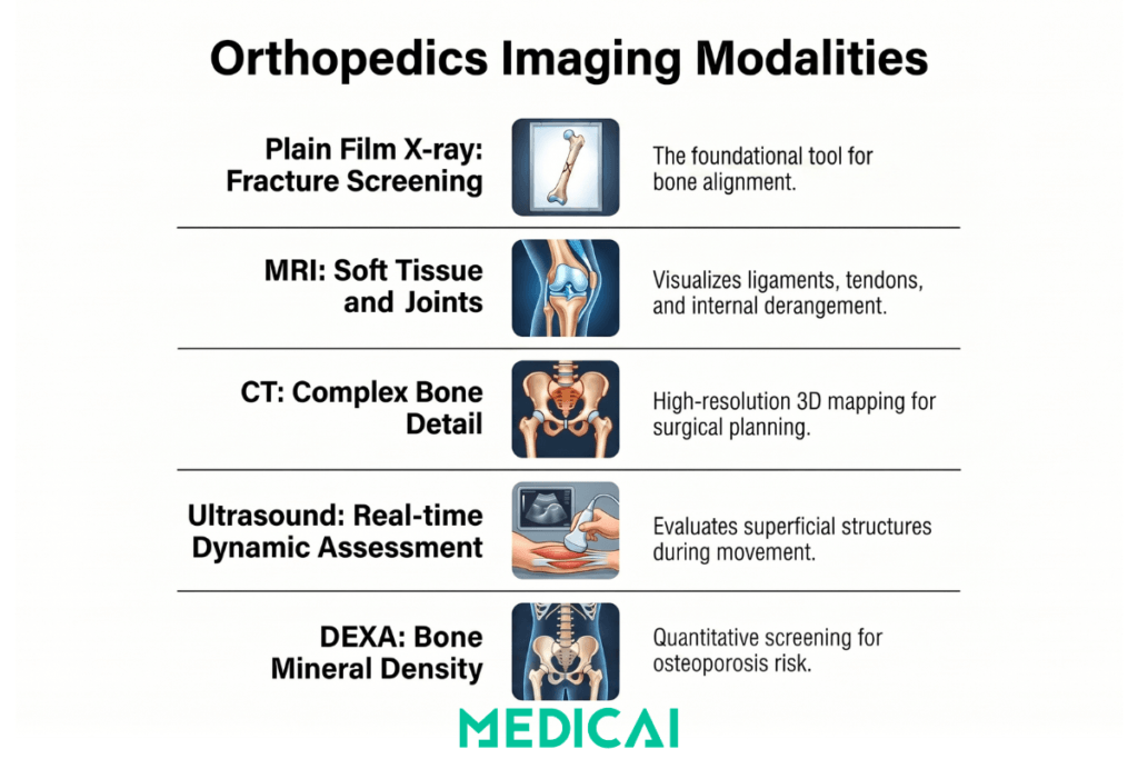
Orthopedic imaging relies on five primary modalities (X-ray, MRI, CT, musculoskeletal ultrasound, and DEXA), and each answers a different question. X-ray and CT show bone. MRI and ultrasound show soft tissue. DEXA measures bone density. Fluoroscopy and nuclear medicine support procedures and hard-to-find injuries. For a broader overview, see our guide to imaging modalities in radiology.
| Modality | Primary orthopedic use | Typical exam time | Effective radiation dose (example) | 2026 Medicare national payment (example) | Main limitation |
|---|---|---|---|---|---|
| X-ray | Fractures, joint alignment, implant position | 5 to 10 minutes | Extremity: under 0.001 mSv. Lumbar spine: 1.4 mSv | $35.74 (shoulder X-ray, CPT 73030) | No soft tissue detail |
| MRI | Ligaments, tendons, cartilage, menisci, labrum, marrow, spinal discs and nerves | 30 to 60 minutes | None (no ionizing radiation) | $204.41 (knee MRI without contrast, CPT 73721) | Cost, scan time, some implants, claustrophobia |
| CT | Complex fractures, bony surgical planning, 3D reconstruction | Usually within 30 minutes | Spine: 8.8 mSv. Abdomen and pelvis: 7.7 mSv | $129.93 (lumbar spine CT without contrast, CPT 72131) | Radiation dose, limited soft tissue contrast |
| Musculoskeletal ultrasound | Tendons, nerves, effusions, guided injections | About 30 minutes | None | $53.78 (complete joint ultrasound, CPT 76881) | Operator-dependent, limited deep views |
| DEXA | Bone density, osteoporosis, fracture risk | 10 to 30 minutes | 0.001 mSv | $39.41 (axial DXA, CPT 77080) | Measures density, not anatomy |
Sources: exam times and doses from RadiologyInfo.org (radiation dose, bone X-ray, CT, MRI, ultrasound, DEXA). Payments are based on the 2026 Medicare Physician Fee Schedule national global amounts (CMS). They are not cash prices. Hospital facility fees, commercial insurance rates, and self-pay prices are usually higher and vary by location. For reference, the average person in the US receives about 3 mSv a year from natural background radiation.
Plain film X-ray (radiography)
X-ray is the foundation of orthopedic imaging and is usually the first study ordered. It detects and classifies fractures, shows joint alignment for replacement planning, and confirms implant position after surgery. Weight-bearing views show lower-limb alignment under load, and spine films assess scoliosis. Orthopedic surgeons read most plain films themselves. X-ray does not show cartilage, ligaments, tendons, or menisci, and subtle or nondisplaced fractures can be missed early. Radiography can also be read with AI support. Gleamer BoneView, for example, received FDA 510(k) clearance in 2022 as a concurrent reading aid for fractures on adult radiographs. For whole-spine and lower-limb alignment, low-dose biplanar systems such as EOSedge are FDA-cleared for general radiographic exams.
Magnetic resonance imaging (MRI)
MRI is the reference standard for soft tissue and internal joint structures, including cartilage, ligaments, tendons, menisci, labrum, muscle, bone marrow, and nerves. Common orthopedic uses include meniscus and ACL tears, rotator cuff and labral injuries, hip labral tears and femoroacetabular impingement, and disc herniation or spinal stenosis. MRI uses no ionizing radiation. 3 Tesla scanners provide higher resolution than 1.5-Tesla scanners, and dedicated extremity coils improve images of the hand, wrist, foot, and ankle. Gadolinium contrast is used for post-operative, infection, and tumor questions; see MRI with contrast. MRI costs more and takes longer than X-ray or CT, and some implanted devices or claustrophobia can rule it out.
Computed tomography (CT)
CT gives high-resolution bone detail and 3D reconstruction, so it is used for complex fractures and bony surgical planning. Typical cases include tibial plateau, calcaneus, acetabulum, and distal radius fractures, revision joint replacement templating, spinal fusion and pedicle screw planning, and bone tumors. CT exposes patients to more radiation than X-ray (about 8.8 mSv for a spine CT, roughly equivalent to three years of natural background radiation), which limits routine use in children and young adults. Soft-tissue contrast is weaker than in MRI. For a side-by-side comparison, see MRI vs. CT.
Musculoskeletal ultrasound
Musculoskeletal ultrasound visualizes superficial soft tissues in real time, including during movement or under load. It is used for the rotator cuff, Achilles and other tendons, tennis elbow and other tendinopathies, peripheral nerves (carpal tunnel, ulnar nerve), and joint effusions. It also guides corticosteroid, platelet-rich plasma, and hyaluronic acid injections. It can be done at the point of care during an office visit, uses no radiation, and has no implant or claustrophobia restrictions. Image quality depends on the operator, and ultrasound cannot see inside bone or evaluate deep structures well.
Dual-energy X-ray absorptiometry (DEXA or DXA)
DEXA measures bone mineral density to screen for and monitor osteoporosis, estimate fracture risk (with tools such as FRAX), and check bone quality before joint replacement or spinal fusion. Results are reported as T-scores and Z-scores. Using WHO criteria, a T-score of -2.5 or lower indicates osteoporosis, and a score between -1.0 and -2.5 indicates osteopenia (RadiologyInfo). The dose is very low, about 0.001 mSv, roughly one-hundredth of a chest X-ray. DEXA measures density only and does not show anatomic injury.
Adjunct modalities: fluoroscopy and nuclear medicine
Fluoroscopy provides real-time X-ray during procedures. C-arm units guide fracture reduction and implant placement in the operating room, and fluoroscopy also guides vertebroplasty, kyphoplasty, injections, and arthrography. Nuclear medicine bone scans detect increased bone turnover. They help find stress fractures, occult fractures, infection, and metastatic disease when X-rays are negative but clinical suspicion is high. PET-CT is used in orthopedic oncology for staging and treatment response.
Clinical Use Cases by Body Region and Subspecialty
Orthopedic imaging choices vary by body region and subspecialty, as each area has its own common injuries and first-line study. The patterns below reflect common practice. Individual choices follow the clinical exam and guidance such as the ACR Appropriateness Criteria.
Sports medicine imaging
Sports medicine imaging serves younger, active patients, where return-to-play decisions depend on fast, accurate diagnosis. The usual sequence:
- X-ray first, to rule out fracture.
- MRI when the exam suggests a soft tissue injury.
- Ultrasound for dynamic assessment and guided injections at the point of care.
Common questions include ACL and other knee ligament injuries, rotator cuff and labral tears, ankle sprains, stress fractures in endurance athletes, and tendinopathy.
Joint replacement and arthroplasty imaging
Joint replacement imaging spans the whole life of the implant. Before surgery, weight-bearing X-rays guide component sizing and alignment through digital templating. CT is added for complex revisions or major deformity, and MRI in selected cases. After surgery, X-rays confirm implant position, and surveillance films track alignment and the bone-implant interface. Targeted imaging follows if pain, instability, or a complication appears. Because implants are expected to last many years (the AAOS reports that more than 90% of modern total knee replacements still function well 15 years after surgery), the full imaging history must remain available for comparison.
Spine imaging
Spine imaging covers the cervical, thoracic, lumbar, and sacral spine for degenerative disease, disc problems, deformity (scoliosis, kyphosis), trauma, infection, tumors, and post-operative checks.
- X-ray: the first study for trauma, alignment, and post-operative follow-up. Flexion-extension views assess instability.
- MRI: the reference standard for discs and nerves, including herniation, stenosis, and nerve root compression.
- CT: bony detail for fusion and deformity planning, including pedicle anatomy and bone quality. CT myelography is used when MRI is not possible.
Spine practices build up large, multi-modality image histories for each patient.
Hand, wrist, and upper extremity
Hand and wrist imaging requires high resolution to visualize small bones, ligaments, tendons, and nerves.
- X-ray: specialized views (scaphoid, oblique, ulnar deviation) for fractures.
- MRI: dedicated extremity scanners or small coils for intrinsic wrist ligaments and tendons.
- Ultrasound: widely used here because the structures are superficial. Uses include carpal tunnel syndrome, De Quervain’s tenosynovitis, trigger finger, and guided tendon sheath injections.
Foot and ankle
Foot and ankle imaging starts with weight-bearing X-rays, which are essential for alignment and biomechanics. MRI covers soft tissue and joint injuries. CT covers complex fractures and surgical planning. Ultrasound evaluates the Achilles and peroneal tendons and the plantar fascia. Stress fractures are a known challenge: early X-rays are often negative, so MRI or a bone scan is used when suspicion stays high.
Pediatric orthopedics
Pediatric orthopedic imaging weighs radiation exposure more heavily than adult imaging, because children are more sensitive to it and have a lifetime of exposure ahead. Growing skeletons also show growth plates and ossification centers that are absent in adults. Age-specific conditions include developmental dysplasia of the hip, slipped capital femoral epiphysis, Legg-Calvé-Perthes disease, and idiopathic scoliosis.
- X-ray: remains the foundation.
- Ultrasound: central to infant hip screening, with no radiation.
- MRI: used more often to avoid radiation when it can answer the question.
In-office orthopedic imaging: equipment and setup
In-office orthopedic imaging means a practice owns and runs its own imaging equipment, so patients can be imaged and diagnosed in the same visit. Practices often start with X-ray and add ultrasound, DEXA, or extremity MRI as volume grows. Each step adds equipment, space, staffing, regulatory, and IT requirements.
Which modalities orthopedic practices bring in-house
The modalities orthopedic practices bring in-house are those that answer the most frequent questions with the lowest setup burden.
- X-ray: usually the first in-house modality, because nearly every fracture, alignment, and post-operative visit needs one.
- Musculoskeletal ultrasound: point-of-care diagnosis and guided injections using a compact unit, with no radiation.
- DEXA: for practices managing osteoporosis or bone health before surgery.
- Extremity MRI: dedicated units image the limbs and joints in a smaller footprint than a whole-body scanner. Esaote’s G-scan Brio received FDA 510(k) clearance in 2012 for limbs, joints, and spine.
- Weight-bearing CT: cone-beam systems image the lower extremities under load. CurveBeam’s HiRise received FDA 510(k) clearance in 2020 for upper and lower extremity and pelvis imaging.
What an in-office imaging setup needs
An in-office orthopedic imaging setup needs five things beyond the equipment itself.
- State registration and inspection. X-ray machines must be registered with the state radiation control program. California, for example, requires registration within 30 days of acquiring a radiation machine, and Florida inspects registered machines on a set schedule. Check your own state’s rules for shielding, operator credentials, and inspections.
- Safe siting for MRI. MRI suites follow the ACR Manual on MR Safety, which divides the site into access-controlled zones around the magnet room.
- A plan for who reads the studies. Orthopedic surgeons can interpret their own images. Medicare pays the professional component of imaging regardless of the physician’s specialty, and requires a written report. Many practices still send MRI and complex CT to musculoskeletal radiologists.
- Image storage and sharing. Studies need a DICOM archive that keeps years of priors for comparison, shares images with referring physicians and patients, and supports templating. A cloud PACS avoids on-site servers. See how to compare cloud PACS vendors.
- Billing compliance. Medicare’s physician self-referral (Stark) rules cover imaging. In-house imaging generally relies on the in-office ancillary services exception. For MRI, CT, and PET, that exception requires providing patients with a written list of at least 5 other suppliers within 25 miles. Review your setup with healthcare counsel.
In-house vs. outsourced imaging: the tradeoffs
The choice between in-house and outsourced imaging comes down to patient convenience and control versus capital cost and compliance work.
| Factor | In-house imaging | Outsourced imaging |
|---|---|---|
| Patient experience | Same-visit imaging and diagnosis | Separate appointment at an imaging center |
| Upfront cost | Equipment, space, and shielding | None |
| Staffing | Technologists, plus physicist and service contracts | Handled by the imaging center |
| Regulatory work | State registration, inspections, Stark compliance | Mostly the imaging center’s responsibility |
| Interpretation | In-house or contracted radiologists | The imaging center’s radiologists |
| Image access | Images in your own PACS immediately | Depends on the center’s image sharing |
The Orthopedic Imaging Workflow
The orthopedic imaging workflow runs in six stages, from the clinical exam to follow-up years after surgery. Each handoff is a point at which images or information can be delayed or lost.
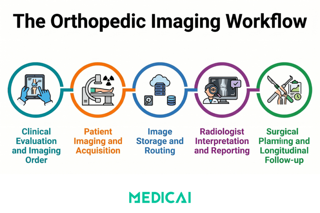
- Clinical evaluation and imaging order. The clinician takes a history, examines the patient, and orders a study that names the body part, the clinical question, and the protocol. In most practices, the order is sent electronically from the EHR to the imaging system, typically as an HL7 order message.
- Image acquisition. A technologist images the patient using a protocol for that body part and question. Image quality depends on positioning and modality settings: exposure factors for X-ray and CT, pulse sequences for MRI, and transducer choice for ultrasound.
- Storage and worklist routing. The modality sends images in DICOM format to the PACS. The PACS stores them and routes them to a reading worklist, sometimes matched to a subspecialist (for example, sports MRI to a musculoskeletal radiologist).
- Interpretation. A radiologist or the treating physician reviews the images in light of the clinical question and writes a report. Structured radiology reporting keeps orthopedic reports consistent and easy to compare. Musculoskeletal radiologists often read complex sports, tumor, and post-operative cases.
- Surgical planning and templating. For surgical patients, the surgeon uses calibrated images to size components and plan alignment, saves the plan, and refers to it in the operating room. Our guide to digital orthopedic templating covers this step in detail.
- Post-operative imaging and long-term comparison. Post-operative images are compared with pre-operative ones to confirm the result, spot complications, and track healing. For joint replacements, this comparison can continue for more than 15 years, so the archive has to keep every prior study accessible.
Orthopedic PACS and templating
An orthopedic PACS stores and displays imaging like any PACS. It also needs tools built for orthopedic surgery:
- Calibrated digital templating for joint replacement sizing
- Side-by-side comparison of X-ray, CT, and MRI across years of priors
- Access for surgeons in the clinic, at home, and in the operating room
Medicai’s cloud-native orthopedic PACS stores DICOM data on Microsoft Azure. It includes a zero-footprint browser viewer with orthopedic tools and integrated templating, so a surgeon can template in the office and pull up the plan in the operating room. For requirements, templating methods, and evaluation criteria, read our orthopedic PACS and templating guide.
Orthopedic Imaging Modalities Compared at a Glance
| Modality | Primary clinical use | Strengths | Limitations | Typical cost (US) |
|---|---|---|---|---|
| Plain film X-ray (radiography) | Initial fracture detection, joint alignment, post-operative implant assessment, skeletal survey | Fast acquisition (2 to 3 minutes), widely available, low cost, immediate interpretation by orthopedic surgeons for most findings | No soft tissue visualisation; subtle fractures may not appear; two-dimensional projection of three-dimensional anatomy | $50 to $250 per study |
| MRI (magnetic resonance imaging) | Soft tissue and internal joint structure assessment: ligaments, tendons, cartilage, menisci, labrum, neural structures | Superior soft tissue contrast, no ionising radiation, multiplanar imaging, three-dimensional reconstruction | Higher cost than other modalities, longer acquisition time (30 to 60 minutes), limited availability in some settings, contraindications for some implanted devices and patient claustrophobia | $1,000 to $4,000 per study |
| CT (computed tomography) | Complex fracture characterisation, pre-operative bony surgical planning, three-dimensional reconstruction | High-resolution bone detail, fast acquisition (under 2 minutes), multiplanar reconstruction, three-dimensional rendering | Meaningful radiation dose (limits use in paediatric and younger adult populations), limited soft tissue contrast compared to MRI | $500 to $2,500 per study |
| Musculoskeletal ultrasound | Superficial soft tissue evaluation, dynamic assessment, image-guided injections, point-of-care imaging | Real-time dynamic imaging, point-of-care availability, no radiation, lower cost, no contraindications | Operator-dependent quality, limited deep structure visualisation, limited bone interior assessment, narrower field of view | $200 to $800 per study |
| DEXA (bone densitometry) | Osteoporosis screening, fracture risk assessment, monitoring of osteoporosis treatment response | Quantitative bone density measurement, very low radiation dose, standardised T-score and Z-score reporting | Does not visualise anatomic pathology; specific to bone density assessment rather than general orthopedic imaging | $100 to $300 per study |
Frequently Asked Questions
Orthopedic doctors use five main scans. X-ray is usually first, for fractures and alignment. MRI shows soft tissue such as ligaments, tendons, cartilage, and menisci. CT gives detailed bone images for complex fractures and surgical planning. Musculoskeletal ultrasound evaluates tendons and nerves and guides injections. DEXA measures bone density. Fluoroscopy and bone scans are used in specific situations.
Yes. Orthopedic surgeons routinely order MRIs when the exam suggests a soft tissue or joint injury. They can also interpret their own imaging, and Medicare pays the professional component regardless of the physician’s specialty, with a written report required. Many practices still send MRIs to musculoskeletal radiologists. Facilities seeking ACR MRI accreditation must meet its training and case requirements for any interpreting physician.
Cost depends on the modality, setting, and insurance. As a reference point, 2026 Medicare national physician fee schedule payments are about $36 for a shoulder X-ray, $54 for a complete joint ultrasound, $39 for a DEXA scan, $130 for a lumbar spine CT, and $204 for a knee MRI without contrast. Hospital facility fees, commercial rates, and self-pay prices are usually higher and vary by location.
Sports injuries usually start with an X-ray to rule out a fracture. MRI follows when a soft tissue injury is suspected, such as an ACL, meniscus, rotator cuff, or labral tear. Ultrasound is used at the point of care for tendons and guided injections. For suspected stress fractures, MRI or a bone scan is used when early X-rays are negative.
Imaging devices send studies to a PACS in DICOM format, where they are stored, read, and shared. An orthopedic PACS adds calibrated digital templating for implant sizing, side-by-side comparison with prior studies, and access for surgeons in the clinic and operating room. The same archive keeps pre-operative and post-operative images for comparison over the life of an implant.
Medicai provides a cloud-native orthopedic PACS with integrated templating, so practices can store, read, plan, and compare orthopedic imaging in one place without on-site servers. To see it with your own studies, explore Medicai for orthopedics or book a demo.


Related Articles

Lets get in touch!
Learn more about how Medicai can help you strengthen your practice and improve your patients’ experience. Ready to start your Journey?
Book A Free Demo