The Future of Medical Imaging: AI, Cloud-Native Infrastructure, and Workflow Transformation Through 2030

The future of medical imaging is being shaped by three converging forces: artificial intelligence moving from pilot to production at scale, cloud-native infrastructure replacing legacy on-premise PACS as the default deployment model, and workflow transformation driven by teleradiology, multi-site practice consolidation, and remote reading. The FDA’s own AI/ML‑enabled medical device list shows that this technology is well past the experimental stage. By late 2024, the cumulative number of authorized AI‑enabled devices had already exceeded 1,000, and by the end of 2025 the total reached about 1,450 across radiology, cardiology, and other specialties. In 2025 alone, the FDA cleared nearly 300 AI‑enabled devices, underscoring that AI in imaging and signal analysis is now an operational reality rather than a distant promise.
The next five years through 2030 will see these forces reshape not just the technology of medical imaging but the organizational structures, regulatory frameworks, and clinical workflows that surround it.

Where medical imaging stands in 2026
The global medical imaging market was valued at roughly $44–46 billion in 2025 and is expected to grow at about 5–6% per year, driven by aging populations, the rising burden of chronic diseases, and an operational shift toward earlier diagnosis through screening and preventive programs. Imaging volume growth has outpaced radiologist workforce growth in most developed markets, creating sustained pressure to extract more diagnostic throughput from the same reading capacity. This pressure is the primary commercial driver behind every trend covered below.
Modality distribution in 2026 remains dominated by X-ray and CT in absolute volume, with MRI growing faster than either, ultrasound holding steady as the workhorse of point-of-care imaging, and PET expanding in oncology and neurology applications. Mammography screening volumes are stable in markets with mature programs and growing in markets adopting them. Specialty imaging (dental CBCT, ophthalmology OCT, cardiology imaging) is growing faster than general radiology, reflecting the broader shift toward specialty-specific imaging workflows.
The infrastructure layer is in the middle of a generational transition. On-premise PACS still dominates legacy hospital deployments, particularly in academic medical centers with significant existing infrastructure investments. Cloud-native imaging is the default for new deployments, imaging center networks, and any practice considering CBCT or OCT for the first time. The crossover point is approaching faster than industry analysts predicted in 2020.
Imaging informatics standards are also shifting. DICOM remains the universal protocol for medical images, but its implementation is migrating from classic DIMSE network protocols (C-FIND, C-MOVE, C-STORE) to DICOMweb (QIDO-RS, WADO-RS, STOW-RS) running over HTTPS. HL7 and FHIR carry the integration between imaging and the broader EHR ecosystem. These are not new standards, but their adoption patterns are changing how imaging systems get architected.
AI is rewriting the radiology workflow
Artificial intelligence has clearly crossed the threshold from research promise to operational reality in medical imaging. By late 2024, the FDA had authorized over 1,000 AI/ML‑enabled medical devices, and by the end of 2025 that total reached roughly 1,450, with radiology accounting for about three‑quarters of all authorizations. In recent years, the number of new AI/ML clearances has grown to several hundred per year, including close to 300 devices in 2025 alone. Most of these tools are classified as clinical decision support or assistive systems rather than fully autonomous interpreters, meaning the radiologist retains ultimate diagnostic responsibility while the AI executes specific tasks—such as triage, detection, or quantification—within the workflow.
AI in clinical reading workflows has matured along three distinct lines. Triage AI flags suspected emergent findings before the radiologist opens the study, prioritizing the worklist by suspected severity rather than chronological arrival. Tools like Viz.ai for large vessel occlusion stroke, Aidoc for intracranial hemorrhage and pulmonary embolism, and Rapid AI for stroke imaging are deployed in thousands of hospitals as of 2026. The clinical impact is measurable: door-to-needle times for stroke have meaningfully shortened at facilities that integrate triage AI with their stroke protocols.
Detection AI assists the radiologist during the read itself. Lunit INSIGHT and Annalise.ai operate on chest X-rays to flag suspected nodules, consolidation, and pneumothorax. iCAD ProFound AI and ScreenPoint Transpara analyze mammography studies to flag suspicious lesions. The pattern across detection AI is consistent: the tool surfaces findings for the radiologist to verify or dismiss, with the radiologist remaining the final decision authority. Sensitivity improvements without unacceptable specificity tradeoffs have been the operational test these tools must pass.
Quantitative measurement AI handles the structured data extraction that humans do slowly and inconsistently. Cardiac CT calcium scoring, brain volumetrics for tracking neurodegenerative disease, liver iron quantification, and tumor burden measurement in oncology are all being automated. These tools don’t replace radiologist interpretation, but they eliminate the manual measurement steps that have historically consumed reading time and introduced inter-observer variability.
AI in the imaging chain upstream of reading is the area expanding fastest in 2026. Deep learning image reconstruction has moved from research to routine clinical use. GE Healthcare’s AIR Recon DL, Siemens Healthineers’ Deep Resolve, and AIRS Medical’s SwiftMR accelerate MRI acquisition while preserving or improving image quality. Philips and Canon offer similar capabilities. The operational impact is significant: shorter scan times mean higher patient throughput from the same MRI suite, reducing the access bottleneck that constrains MRI volume in most health systems. Protocol optimization AI handles automated parameter selection for CT and MRI based on the clinical question and patient characteristics, reducing operator variability and improving consistency.
The regulatory landscape is evolving in parallel with the clinical deployment. The FDA’s De Novo and 510(k) pathways have absorbed most of the AI/ML clearances to date, with the FDA’s predetermined change control plan (PCCP) framework introduced to handle AI tools that learn and update post-clearance. The EU’s Medical Device Regulation and the AI Act create overlapping requirements for AI tools deployed in European healthcare. Compliance complexity is now a material factor in AI vendor evaluation, not just a checkbox.
The question every radiologist asks deserves a direct answer. Current evidence does not support AI replacing radiologists. AI is reshaping radiologists’ workflows by handling triage, detection, and quantitative measurement, freeing radiologists to focus on complex reasoning, clinical correlation, and patient communication. Workforce demand for radiologists remains strong through 2030, though the role is evolving toward AI-augmented practice. For deeper coverage of AI’s clinical applications, see Medicai’s AI in radiology overview.
Cloud-native infrastructure becomes the new baseline
The shift from on-premise PACS to cloud-based imaging is the architectural decision driving more capital allocation in medical imaging through 2030 than any other infrastructure choice. Cloud-native imaging platforms now serve as the default for new deployments, imaging center networks, and multi-site practice consolidation, while existing on-premise installations are migrating in waves as hardware refresh cycles force the decision.
The driver is the file-size economics of modern imaging. Optical coherence tomography in ophthalmology produces 100-500 MB per scan. Cone-beam CT in dentistry produces files within similar ranges. High-resolution mammography, photon-counting CT, and wide-bore MRI all push file sizes in the same direction. The cumulative storage burden across a multi-site practice or hospital network makes per-site on-premise archives operationally impractical and economically wasteful.
Vendor-neutral archive (VNA) architecture has emerged as the structural answer to a related problem: the proliferation of vendor-specific imaging silos that lock each modality’s data into the proprietary archives of their acquisition software. VNA decouples the imaging storage layer from the PACS reading software, allowing a single archive to serve multiple imaging vendors, multiple reading workflows, and multiple sites. The 2026 Best in KLAS VNA winner is AGFA HealthCare Enterprise Imaging at 89.8 percent satisfaction, marking its second consecutive year at the top.
The economic case for cloud-native deployment has matured beyond simple CapEx-to-OpEx framing. Cloud platforms eliminate hardware refresh cycles, IT staff overhead for storage management, and the operational risk of running aging infrastructure past its useful life. Elastic storage scales automatically as study volume grows, with hot-warm-cold tiering managing the cost curve as studies age out of active access. Geo-redundant replication provides disaster recovery without the operational discipline that on-premise backup strategies depend on.
The compliance and security pattern has also matured. Cloud platforms with SOC 2 Type II audits, HIPAA Business Associate Agreement availability, GDPR compliance for European deployments, and ISO 27001 certifications have become baseline expectations rather than premium differentiators. The security pattern that worried IT teams about cloud in 2018 has reversed: cloud platforms now typically have stronger security postures than the on-premise systems they replace, because cloud vendors invest in security at a scale individual hospitals cannot match.
Workflow transformation: teleradiology, multi-site practices, and remote reading
Radiology workflow patterns are undergoing the most significant restructuring since the original transition from film to digital. Three concurrent shifts are reshaping how reading actually happens day to day.
Teleradiology has expanded far beyond its original after-hours coverage role. The traditional nighthawk model, which emerged in the early 2000s, addressed overnight emergency imaging when in-house radiologists were unavailable. The 2026 teleradiology landscape covers subspecialty routing during daytime hours (pediatric neuroradiology, MSK, breast imaging routed to fellowship-trained specialists regardless of geography), daytime overflow management for high-volume facilities, and geographic load balancing across multi-state practice networks. Roughly 70 percent of US hospitals use after-hours teleradiology for at least some portion of overnight coverage as of 2026.
Multi-site practice patterns have transformed the architectural requirements for imaging infrastructure. Single-clinic practices operated each location as an island, with imaging data accessible only locally. Multi-site practices require centralized worklists, floating clinicians who can access patient imaging from any clinic without a VPN setup, and referring-specialist workflows that connect general practices with subspecialty consultants in seconds rather than days. Cloud-native imaging architecture is the operational prerequisite for these workflow patterns; on-premise PACS at each location actively blocks them.
Remote-first reading is now permanent rather than the pandemic-era exception it started as. Radiologists reading from home offices, distributed reading centers, and shared subspecialty hubs are the default operational pattern at most teleradiology groups and a growing share of in-house radiology departments. The technology stack supporting this includes browser-based diagnostic viewers, AI triage that prioritizes worklists across distributed reading teams, and a cloud-native PACS architecture that eliminates the VPN dependency required by the legacy remote-reading model.
The integration point that ties these three shifts together is AI triage routing. A study acquired at one facility, flagged by AI as a suspected stroke, is routed to the next available neuroradiologist with the appropriate credentials and licensure, regardless of the radiologist’s physical location. The legacy workflow of manual study assignment, geographic constraint, and chronological worklist sorting is being replaced by AI-curated, multi-site, subspecialty-routed reading.
DICOM and imaging standards in the AI era
DICOM remains the universal protocol for medical imaging in 2026 and through 2030, but its implementation patterns are shifting in ways that affect every layer of the imaging stack.
The migration from classic DIMSE protocols (C-FIND, C-MOVE, C-STORE running over dedicated TCP ports) to DICOMweb (QIDO-RS, WADO-RS, STOW-RS running over HTTPS) is accelerating. DICOMweb’s structural advantages are operational: it operates over standard HTTPS rather than requiring dedicated network ports, traverses firewalls cleanly, supports REST-based queries that integrate naturally with cloud architectures, and enables browser-based clients without the network engineering overhead that DIMSE requires. Legacy DIMSE remains common in established systems but is being deprecated in new deployments.
DICOM Structured Reporting (DICOM SR) has emerged as the carrier for AI-generated structured findings. Rather than embedding AI outputs in free-text radiologist reports, DICOM SR provides a structured data envelope that carries quantitative measurements, anatomical locations, and AI confidence scores in a machine-readable format that downstream systems can consume programmatically. This standard has existed since the 1990s but is finally seeing widespread implementation as AI outputs require structured carriage rather than narrative description.
The relationship between DICOM and FHIR is increasingly important. DICOM handles imaging-specific data (pixels, metadata, structured reports); FHIR handles general healthcare data exchange (patient demographics, encounters, observations, allergies, medications). FHIRcast and similar integration frameworks bridge the two, enabling launch-in-context viewing, in which an EHR pulls imaging from a separate PACS with proper patient and study context. The two standards are not competitors but complementary layers of the healthcare interoperability stack.
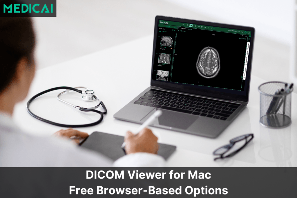
Browser-based DICOM viewers have become the default for new deployments. Open-source toolkits like Cornerstone, OHIF, and dwv power most modern web-based viewers, eliminating the per-workstation thick-client installation that legacy radiology workstations required. The diagnostic-grade browser viewer is no longer a novelty but a category, with FDA-cleared web viewers competing on tooling depth, modality coverage, and integration capability. Medicai’s own DICOM viewer is one example of this category; see our best DICOM viewers comparison for the broader landscape.
What radiology practices should do now to prepare for 2030
Four moves separate practices that will be operationally well-positioned in 2030 from those that will be playing catch-up.
Prioritize cloud-native imaging infrastructure for new deployments and plan migration for legacy on-premise. New practice deployments, new imaging centers, and any practice considering CBCT, OCT, or other large-file modalities should default to cloud-native architecture. For existing on-premise PACS deployments, plan the migration timeline around the next hardware refresh cycle rather than reactively when the current infrastructure fails. The migration is operationally significant but increasingly routine; the practices delaying it are accumulating technical debt that compounds with each passing year.
Adopt high-ROI AI tools rather than betting on speculative AI. The AI tools with proven clinical and operational ROI in 2026 are concentrated in triage (stroke, intracranial hemorrhage, pulmonary embolism), detection (mammography, chest X-ray), and quantitative measurement (cardiac calcium scoring, brain volumetrics). These tools have FDA clearance, established deployment patterns, and measurable outcomes data. Speculative AI tools that promise broader autonomous interpretation should be evaluated carefully against actual clearance status, evidence of real-world deployment, and the regulatory pathway for the specific use case.
Invest in radiologist skills that complement AI rather than skills AI replaces. Subspecialty depth, AI tool curation and integration, clinical reasoning under diagnostic uncertainty, and patient and referring-clinician communication are the skills appreciating in value as AI handles more of the routine detection and measurement work. Skills that AI handles well (basic detection, quantitative measurement, routine reading throughput) are becoming less valuable as differentiators, even though they remain part of the radiologist’s role.
Evaluate vendors on architectural fit rather than feature lists. Vendor-neutral architecture, support for open standards (DICOMweb, FHIR, HL7), AI orchestration capability, and platforms that scale across single-site and multi-site deployments are the architectural patterns that hold up through the next decade. Vendors locked into proprietary formats, dedicated thick-client viewers, or single-modality silos are increasingly out of step with the industry’s direction. For practices evaluating modern cloud-native imaging platforms, Medicai provides one example of this architectural pattern.
Looking beyond 2030
The trends covered above are not the end state but a waypoint. Several developments visible in 2026 will shape medical imaging through 2030-2035 in ways that are harder to predict precisely but worth tracking.
Multimodal AI integrating imaging with genomics, clinical notes, laboratory results, and pathology data represents the next frontier beyond imaging-only AI. The early research is concentrated in oncology, where treatment decisions depend on integrating imaging, molecular markers, and clinical context. Production deployment of multimodal AI is probably 5-7 years away for routine clinical use, but the architectural decisions made in 2026 will determine whether health systems are positioned to adopt it when it arrives.
AI-generated synthetic imaging for training, protocol development, and education is moving from research to early production. Synthetic CT and MRI generated by diffusion models can augment training datasets for AI development, support protocol optimization without patient scanning, and provide standardized cases for radiology education. The clinical use case is more nuanced; synthetic imaging is unlikely to replace patient imaging for diagnosis, but supporting roles in workflow optimization are expanding.
Theranostics and the integration of imaging with treatment in oncology are the areas where imaging is becoming most operationally entwined with clinical decision-making. PET-guided molecular therapy planning, image-guided radioembolization, and prostate-specific membrane antigen PET for prostate cancer treatment selection are examples in which imaging is no longer just diagnostic but is operationally integrated into the treatment workflow.
https://blog.medicai.io/en/radiology-modalities/Continued specialization within radiology, toward subspecialty fellowship training and AI-augmented practice models, is the workforce trend that compounds the technology trends above. The general radiologist reading across all modalities and anatomical regions remains the dominant model in 2026, but the trajectory is toward subspecialty depth, augmented by AI tools tuned to specific clinical questions.
Frequently asked questions about the future of medical imaging
The future of medical imaging through 2030 is being shaped by three converging forces: artificial intelligence augmenting radiologist workflows at scale, cloud-native infrastructure replacing legacy on-premise PACS, and workflow transformation through teleradiology, multi-site practice consolidation, and remote reading. The FDA has cleared over 700 AI/ML medical devices as of 2024, most in radiology, signaling that AI has crossed from clinical trial into operational reality.
AI is changing medical imaging at three points in the workflow: image acquisition (deep learning reconstruction, accelerated MRI, motion correction), clinical reading (triage AI for stroke and emergency findings, detection AI for mammography and chest X-ray, quantitative measurement tools), and reporting (auto-populated structured findings). Current AI is augmentation rather than autonomous interpretation, with radiologists retaining final diagnostic responsibility.
Current evidence does not support AI replacing radiologists. The 700+ FDA-cleared AI/ML medical devices are categorized as decision support tools, not autonomous interpretation. AI is reshaping radiologist workflows by handling triage, detection, and quantitative measurement, freeing radiologists for complex reasoning, clinical correlation, and patient communication. Workforce demand for radiologists remains strong through 2030, though the role is evolving toward AI-augmented practice.
Cloud computing is shifting medical imaging from on-premise PACS deployments to vendor-managed cloud archives accessible from any authorized location. The shift converts capital expenditure (server hardware, refresh cycles) to operating expenditure (subscription fees), supports multi-site practice consolidation through centralized archives, and enables remote reading without VPN infrastructure. Cloud-native is the default model for new deployments in 2026.
Five trends will dominate medical imaging by 2030: AI integrated into routine reading workflows rather than as a pilot, cloud-native infrastructure as the default deployment model, multimodal AI combining imaging with genomics and clinical data, teleradiology and remote reading as permanent workflow patterns rather than coverage-only options, and continued migration of imaging informatics standards from DICOM-DIMSE to DICOMweb and FHIR.
DICOM remains the universal medical imaging standard, but its implementation is shifting from classic DIMSE network protocols (C-FIND, C-MOVE, C-STORE) to DICOMweb (QIDO-RS, WADO-RS, STOW-RS) running over HTTPS. DICOM Structured Reporting carries AI-generated structured findings. FHIR provides the bridge to broader healthcare data exchange. Browser-based viewers using Cornerstone and OHIF are becoming the new normal for image display.
Radiology workflows through 2030 will see AI triage prioritize worklists by suspected severity rather than chronological arrival, remote reading become permanent rather than coverage-only, multi-site practices consolidate around centralized cloud archives, subspecialty routing direct cases to fellowship-trained readers regardless of geography, and structured reporting replace free-text dictation for AI-augmentable findings. The radiologist’s role evolves toward complex reasoning and AI-curated reading.
Practices should prioritize four moves to prepare for 2030: deploy cloud-native imaging infrastructure for new builds and plan migration for legacy on-premise, adopt high-ROI AI tools (triage, detection) rather than speculative AI, invest in radiologist skills that complement AI (subspecialty depth, AI tool curation, clinical reasoning), and evaluate vendors prioritizing vendor-neutral architecture, open standards, and platforms supporting AI orchestration.

Related Articles
Lets get in touch!
Learn more about how Medicai can help you strengthen your practice and improve your patients’ experience. Ready to start your Journey?
Book A Free Demo