AI in Mammography: How It Works, FDA-Cleared Tools, and What Imaging Centers Need to Deploy It

Artificial intelligence in mammography is the application of machine learning algorithms, primarily deep neural networks trained on large annotated mammographic image datasets, to assist with detecting breast cancer, assessing breast cancer risk, quantifying breast density, triaging screening worklists, and structuring diagnostic reports. AI in mammography does not replace the radiologist. It operates as a computational layer within the breast imaging workflow: the AI processes the digital mammogram or digital breast tomosynthesis (DBT) study, identifies patterns associated with malignancy or elevated risk, and presents its output to the radiologist for verification and clinical interpretation.
The scope of AI in mammography spans five distinct application categories, each addressing a different part of the breast imaging workflow: computer-aided detection of malignant findings, cancer risk prediction models that estimate future cancer probability from current imaging, breast density assessment and BI-RADS density category assignment, screening workflow triage and worklist prioritization, and reporting assistance with BI-RADS template structuring. These categories differ in what they do, where they sit in the clinical workflow, how the FDA regulates them, and how mature they are in clinical deployment. Understanding the distinction between them is the prerequisite for evaluating what AI can realistically deliver in a specific breast imaging practice, and what it cannot.
This guide covers the complete picture: how AI is used across the five mammography application categories, what measurable performance the FDA-cleared products have demonstrated in clinical validation studies, where the limitations and regulatory boundaries sit today, how the different application types compare for breast imaging centers evaluating where to invest, and what PACS infrastructure is required to integrate AI mammography tools into existing screening workflows.
How AI Is Used in Mammography: Five Application Categories
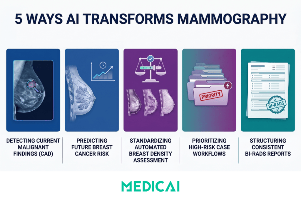
Computer-Aided Detection of Malignant Findings (CAD)
Computer-aided detection in mammography refers to AI systems that identify and mark potential cancers on digital mammograms and digital breast tomosynthesis studies, flagging suspicious masses, microcalcification clusters, architectural distortions, and asymmetries for the radiologist’s review. The current generation of AI CAD is fundamentally different from the older rule-based CAD systems deployed in the 2000s. While legacy CAD relied on hand-engineered features and produced high false-positive rates that reduced radiologist confidence, modern AI CAD uses deep convolutional neural networks trained on millions of annotated mammograms and achieves detection performance closer to that of expert breast radiologists.
Named FDA-cleared products in the AI mammography CAD category include iCAD ProFound AI for both 2D digital mammography and 3D digital breast tomosynthesis, Lunit INSIGHT MMG, ScreenPoint Medical’s Transpara, Therapixel MammoScreen, and Hologic Genius AI Detection. Each of these products operates at the study level, processing the four standard mammographic views (right and left craniocaudal and mediolateral oblique) and producing a per-image overlay highlighting suspicious findings, as well as a case-level malignancy score for the overall study.
The FDA maintains a public database of AI/ML-enabled medical devices. As of 2024, the radiology category accounts for the largest share of FDA-cleared AI devices, and mammography is one of the most active subspecialty areas within that category, with new clearances occurring throughout 2024 and 2025.
Cancer Risk Prediction Models
Cancer risk prediction AI takes a different clinical task from CAD. Where CAD asks “is there cancer in this image now?”, risk prediction asks ‘what is this woman’s probability of developing breast cancer in the next 1 to 5 years, based on imaging features detectable today?’ The clinical value of risk prediction is in screening interval personalization: women at elevated short-term risk benefit from supplemental imaging (breast MRI, contrast-enhanced mammography) or shorter screening intervals, while women at low predicted risk may safely extend their screening interval.
The most recent significant entry in this category is Clairity Breast, which received FDA marketing authorization in 2025 for predicting 5-year breast cancer risk from a standard screening mammogram. The Breast Cancer Research Foundation’s summary of the Clairity Breast clearance describes the approach: rather than detecting visible cancer, the model identifies subtle imaging features that correlate with elevated future cancer risk, producing a 5-year risk score that supplements traditional risk models like the Tyrer-Cuzick and Gail models.
Other risk prediction approaches in active development include the Mirai model from MIT’s Computer Science and AI Laboratory, which predicts 1-year and 5-year breast cancer risk and has been validated across multiple international datasets. The clinical integration challenge for all risk prediction tools is the same: the risk score is only useful if it changes screening management, which requires both clinician adoption and a downstream pathway for women identified as high-risk to receive supplemental imaging.
Breast Density Assessment
Breast density is the proportion of fibroglandular tissue to fatty tissue in the breast, assessed visually by the radiologist and reported using the four BI-RADS density categories (A, almost entirely fatty; B, scattered fibroglandular density; C, heterogeneously dense; D, extremely dense). Density matters clinically for two reasons: dense breast tissue makes mammographic detection of cancer more difficult because fibroglandular tissue and cancer both appear white on mammograms, and dense breast tissue is itself an independent risk factor for breast cancer. As of 2024, the FDA’s updated mammography reporting requirements mandate that breast density be communicated to patients in lay terms, making consistent and accurate density assessment a regulatory and clinical priority.
AI density assessment tools produce automated BI-RADS density category assignments from the screening mammogram, addressing the well-documented interreader variability in visual density assessment. The most established product in this category is Volpara Health’s VolparaDensity, FDA-cleared and widely deployed across US screening practices for objective density measurement. iCAD’s ProFound AI suite includes a density assessment module, and several other AI vendors have integrated automated density assessment into broader mammography AI platforms.
The clinical workflow advantage of AI density assessment is consistency: the same algorithm applied to the same mammogram always produces the same density category, eliminating visit-to-visit variability that can affect manual assessment when different radiologists read the same patient’s serial screenings.
Workflow Triage and Worklist Prioritization
Mammography screening AI can be deployed as a triage layer to process incoming screening studies and reorder the radiologist’s reading worklist based on the AI’s assessment of suspicion. Studies with high case-level malignancy scores are moved to the top of the worklist for earlier radiologist review. Studies with very low malignancy scores may be flagged for first-read confirmation only, depending on the institution’s protocol and the regulatory pathway under which the AI is deployed.
The most operationally significant triage application is in high-volume screening centers that read thousands of mammograms per week, where AI-driven workflow prioritization can reduce time-to-callback for women with suspected cancer and reduce reading time on clearly normal studies. The clinical evidence for triage workflow deployment is most developed in European screening programs operating under regulatory frameworks different from those in the US, where AI is being studied as a replacement for one of the two readers in double-read screening programs.
In the US screening context, AI triage typically operates as a supplementary layer alongside standard single-reader workflows, with the AI’s worklist prioritization and case-level scores informing but not replacing the radiologist’s interpretation. The 2025 RSNA reporting describes ongoing studies on AI-assisted reader workflow in the US screening context.
Reporting Assistance and BI-RADS Structuring
Reporting assistance AI in mammography supports the structured BI-RADS reporting workflow: extracting findings from the radiologist’s dictated text, populating the structured BI-RADS template fields (composition, findings, impression, BI-RADS assessment category, management recommendation), and ensuring that the final report conforms to ACR BI-RADS reporting standards. This is the same reporting AI category covered for radiology generally in the AI in radiology guide, applied specifically to the BI-RADS structured reporting context.
The clinical value of AI-assisted BI-RADS structuring is consistency. BI-RADS classification has documented interreader variability, particularly for the boundary cases between BI-RADS 3 (probably benign) and BI-RADS 4A (low suspicion for malignancy), and between density categories C and D. AI-assisted template population reduces variability by ensuring all required structured fields are completed and that the BI-RADS final assessment category aligns with the findings documented in the report body. The integration architecture for AI-assisted BI-RADS reporting follows the same pattern described in the Medicai structured radiology reporting guide.
What AI Can Do in Mammography: Measurable Benchmarks
Vendor marketing claims about AI mammography performance are abundant. Peer-reviewed clinical validation data are more limited but more useful. The benchmarks below come from published clinical studies and FDA-cleared product validation data, with sources cited.

Cancer detection rate improvement with AI-assisted reading. The most-cited benchmark in the AI mammography evidence base is the iCAD ProFound AI validation, which reported a 9.6% increase in cancer detection rate and a 5.6% reduction in recall rate compared with radiologists reading without AI assistance, published in Radiology. The combination matters clinically: AI tools that increase cancer detection at the cost of higher recall (and therefore more false positives) face significant resistance to clinical adoption. Tools that improve both metrics simultaneously demonstrate genuine clinical utility.
AI-assisted reading versus standard double-read screening. The Mammography Screening with Artificial Intelligence (MASAI) trial, published in The Lancet Oncology, evaluated AI-supported single reading against standard double reading in the Swedish national breast cancer screening program. The AI-supported single read produced a higher cancer detection rate while reducing radiologist reading workload by approximately 44%. The trial is among the strongest pieces of clinical evidence supporting AI-assisted screening workflows in population-based settings.
Cancer detection in dense breasts. AI mammography tools show particular performance gains in dense breasts, where conventional mammography has known sensitivity limitations. The 2024 RSNA reporting on AI breast cancer detection studies documented AI assistance producing measurable increases in cancer detection rates in BI-RADS C and D density categories, where the masking effect of dense fibroglandular tissue most commonly causes missed cancers.
Interval cancer prediction. The 2025 RSNA reporting on AI for interval cancer identification describes ongoing work on AI models that identify women at elevated risk of developing breast cancer between screening rounds. Interval cancers, diagnosed after a negative screening mammogram but before the next scheduled screen, are a known limitation of population screening programs. AI identification of women at elevated interval cancer risk could enable supplemental imaging that captures these otherwise-missed cancers.
Breast density assessment consistency. Automated breast density tools demonstrate near-perfect intra-algorithm consistency (the same mammogram always produces the same density category), addressing the documented interreader variability in visual BI-RADS density assignment. Inter-algorithm consistency across different AI density tools is less perfect, with documented discordance in approximately 10 to 20 percent of cases at the C/D density boundary.
Limitations and Regulatory Status
The capability benchmarks above are real and clinically validated. The limitations that follow are equally real and frequently understated in vendor marketing.
Current Limitations
Validation population versus deployment population mismatch. AI mammography models trained on data from a single demographic, geographic, or screening population may underperform when deployed in other contexts. Models trained predominantly on Northern European screening populations may underperform on US Hispanic or African American populations, where breast density distributions, body habitus, and disease prevalence patterns differ. The generalisability question must be answered for the specific deployment population, not assumed from the original validation study.
Performance variability across mammography hardware. AI mammography tools are trained on images acquired with specific digital mammography systems from specific manufacturers (GE Healthcare, Hologic, Siemens, Fujifilm, Philips). Performance on imaging from other manufacturers, from older equipment generations, or from systems with non-standard acquisition protocols may differ from the validation benchmarks. Before deploying AI, the practice must verify that the AI vendor has validated performance on the specific imaging hardware in use.
Tomosynthesis versus 2D performance differences. Most AI mammography tools have separate validation data for 2D full-field digital mammography and 3D digital breast tomosynthesis. The clinical performance characteristics differ between modalities, and validation on one does not transfer automatically to the other. Practices migrating from 2D screening to DBT screening must verify that their AI tools are validated for DBT input.
Integration friction at deployment. An AI tool that performs well in a controlled validation study may underperform in clinical deployment because its integration with the PACS viewer is poor, its inference time adds latency to the reading workflow, or its output format does not match the radiologist’s reporting structure. The performance gap between published validation and the reality of clinical deployment is consistently underestimated in procurement decisions, whether in mammography or in general radiology AI.
Risk prediction model interpretability. Risk prediction AI tools produce a numerical risk score from a mammogram, but the imaging features the model uses to produce that score are not always interpretable in clinical terms. A woman whose mammogram suggests an elevated 5-year cancer risk reasonably wants to know what about her mammogram drives that prediction. Current risk models offer limited interpretability of the underlying features that drive the risk score, complicating patient communication and shared decision-making.
Regulatory Status
The FDA AI/ML-Enabled Medical Devices database includes all FDA-cleared AI mammography products. Most mammography CAD products operate under 510(k) clearance, demonstrating substantial equivalence to previously cleared CAD predicate devices. The 2025 marketing authorization of Clairity Breast for breast cancer risk prediction represents a different regulatory pathway, reflecting the novel clinical claim of risk prediction (rather than detection) from screening mammography.
The FDA’s mammography regulation framework includes additional requirements specific to breast imaging beyond the general AI/ML SaMD regulatory framework. The Mammography Quality Standards Act (MQSA) governs mammography facility accreditation, radiologist qualifications, and reporting requirements. AI tools used in MQSA-accredited facilities must operate within the MQSA framework, including the 2024 updated mammography reporting requirements that mandate breast density communication to patients.
In the European Union, AI medical devices fall under the Medical Device Regulation (MDR) 2017/745, with most mammography AI tools classified as Class IIa or Class IIb, requiring conformity assessment by a Notified Body. European mammography AI deployment is more advanced in some respects than US deployment, with several large population screening programs (Sweden, Netherlands, Germany) actively studying AI-supported single reading as a replacement for traditional double reading.
AI in Mammography Compared: Five Application Types at a Glance
| AI application type | What it does | Workflow position | Named FDA-cleared products | Clinical maturity and validation |
|---|---|---|---|---|
| Computer-aided detection (CAD) | Identifies and marks suspicious masses, microcalcification clusters, architectural distortions, and asymmetries on 2D digital mammography and digital breast tomosynthesis (DBT) studies for radiologist verification | During image review — AI overlays appear on the PACS viewer alongside the original mammographic images for the radiologist to review and verify | iCAD ProFound AI, Lunit INSIGHT MMG, ScreenPoint Transpara, Therapixel MammoScreen, Hologic Genius AI Detection | Most mature category — iCAD ProFound AI demonstrated 9.6% cancer detection rate increase with 5.6% recall reduction in Radiology validation studies; MASAI trial in The Lancet Oncology demonstrated AI-supported single reading reduced radiologist workload by 44% while increasing detection rate |
| Cancer risk prediction | Predicts 1-year or 5-year breast cancer risk from imaging features detectable in a current screening mammogram, supplementing traditional risk models including Tyrer-Cuzick and Gail | After image review — produces a numerical risk score that informs screening interval personalisation and supplemental imaging decisions (breast MRI, contrast-enhanced mammography) | Clairity Breast (FDA marketing authorisation 2025), Mirai (research-stage, MIT CSAIL) | Emerging — Clairity Breast is the first widely-deployed FDA-cleared risk prediction product; clinical integration pathways still developing as practices establish protocols for routing high-risk patients to supplemental imaging |
| Breast density assessment | Produces automated BI-RADS density category assignment (A almost entirely fatty, B scattered, C heterogeneously dense, D extremely dense) from the screening mammogram, addressing documented interreader variability in visual density assessment | During image review or as a pre-processing step — density category delivered to the radiologist as part of the structured BI-RADS report | VolparaDensity (Volpara Health), iCAD ProFound Density, integrated density modules across most major mammography AI platforms | Established — widely deployed across US screening practices; FDA 2024 updated mammography reporting requirements mandate patient density communication, making consistent density assessment a regulatory as well as clinical priority |
| Workflow triage and worklist prioritisation | Reorders the screening reading worklist based on case-level malignancy scores, prioritising high-suspicion studies for earlier radiologist review and reducing time-to-callback for suspected cancers | Before radiologist review — processes incoming studies within seconds of acquisition and reprioritises the worklist before the radiologist opens any study | iCAD ProFound AI worklist triage, Lunit INSIGHT MMG with workflow integration, ScreenPoint Transpara workflow tools | Operationally significant in high-volume screening centres — clinical evidence strongest in European population screening programmes deploying AI as one of the two readers in double-read screening; US single-reader workflows use triage as supplementary rather than reader replacement |
| Reporting assistance and BI-RADS structuring | Structures the BI-RADS report, populates template fields from dictated text, ensures the BI-RADS final assessment category (0 through 6) aligns with the documented findings in the report body | After image review — processes the radiologist’s findings and dictation into the structured BI-RADS report delivered to the ordering physician’s EHR via HL7 ORU integration | Nuance PowerScribe (radiology-optimised dictation), AI-enhanced BI-RADS template tools integrated into modern cloud PACS platforms | Mature for dictation, emerging for AI-assisted structured BI-RADS field population — same integration architecture as general radiology structured reporting; addresses documented BI-RADS classification interreader variability particularly at the BI-RADS 3/4A boundary and density C/D boundary |
The PACS Infrastructure Requirement for AI Mammography Deployment
AI mammography tools do not operate in isolation. Each of the five application categories described above requires technical integration with the breast imaging practice’s existing PACS and reporting infrastructure to be usable in clinical workflow. The integration requirements are specific, and practices evaluating AI mammography procurement consistently underestimate the infrastructure work involved.
DICOM Mammography Image Storage
Digital mammography images use the DICOM Mammography (DICOM-MG) Information Object Definition, a specific DICOM extension with mammography-specific metadata fields including the standard view positions (right craniocaudal, left craniocaudal, right mediolateral oblique, left mediolateral oblique), implant displacement views, and magnification views. Digital breast tomosynthesis uses the DICOM Breast Tomosynthesis Image Storage SOP class, producing significantly larger studies than 2D mammography (a single DBT screening study typically ranges from 800 MB to 2 GB). Both formats must be preserved with full metadata for AI tools to operate correctly, as the AI uses view-position metadata to determine which images to compare for laterality assessment, year-over-year change detection, and structured reporting.
AI Integration via DICOMweb or Vendor APIs
AI mammography tools integrate with the PACS through three primary architectures. DICOM-based integration with the AI vendor’s on-premise or cloud server, where the PACS forwards mammographic studies via DICOM C-STORE and receives the AI results back via DICOM Structured Reports or DICOM Secondary Capture overlays. DICOMweb integration using WADO-RS for study retrieval, QIDO-RS for worklist queries, and STOW-RS for AI result storage, the standard architecture for cloud-native AI tools. Vendor-specific REST API integration for AI tools that operate outside standard DICOM workflows. The PACS must support whichever integration pattern the AI vendor uses, and the integration must be tested for both 2D mammography and DBT inputs.
Hanging Protocol Configuration for AI Output Display
Mammography reading uses standardized hanging protocols that present the four standard views in a specific arrangement (typically right CC and left CC in the top row, right MLO and left MLO in the bottom row, with prior comparison studies displayed on a second monitor). When AI tools produce overlays or annotations, the hanging protocol must be configured to display the AI output alongside the original images without disrupting the radiologist’s established reading pattern. The work on configuring the hanging protocol for AI integration is consistently underestimated. The general principles of PACS hanging protocol configuration are covered in the Medicai hanging protocols guide.
BI-RADS Reporting Integration
The AI output (case-level malignancy score, per-image findings, density category, risk score) must flow into the structured BI-RADS report delivered to the ordering physician. This requires the PACS reporting platform to accept AI-generated findings as structured input and to format them into the standard BI-RADS report fields. Direct integration between AI tools and reporting platforms is increasingly the standard architecture; older deployments in which AI results are manually transcribed into reports pose workflow risks and productivity bottlenecks.
Medicai’s cloud-native PACS on Microsoft Azure provides the infrastructure layer that supports AI mammography integration: DICOM-MG and DICOM Breast Tomosynthesis storage with full metadata preservation; DICOMweb support for cloud-native AI tool integration; hanging protocol configurability for AI output display; and structured reporting integration with BI-RADS template support. For breast imaging centers evaluating AI mammography deployment, the underlying PACS infrastructure determines whether the AI tool delivers its theoretical performance in the actual reading workflow, or whether integration friction erodes the clinical benefit.
Frequently Asked Questions
AI in mammography is the application of machine learning algorithms, primarily deep neural networks, to assist with detecting breast cancer, assessing breast cancer risk, measuring breast density, triaging screening worklists, and structuring diagnostic BI-RADS reports. AI in mammography does not replace the radiologist. It operates as a computational layer within the breast imaging workflow, processing the digital mammogram and producing outputs (detections, risk scores, density categories, structured report fields) that the radiologist verifies and incorporates into the final clinical interpretation.
Yes, AI is in active clinical use across many US and European breast imaging practices, with adoption accelerating since 2020. The most widely deployed AI applications are computer-aided detection of suspicious findings, automated breast density assessment, and workflow triage of screening worklists. The FDA has cleared over a dozen mammography-specific AI products, with new clearances continuing through 2024 and 2025. The clinical deployment model is consistently radiologist-in-the-loop: the AI produces findings, the radiologist reviews and verifies them, and the radiologist remains responsible for the final diagnostic interpretation and BI-RADS assessment category.
The clinical and research consensus is that AI augments rather than replaces breast radiologists. AI excels at high-volume, narrow-task pattern recognition on screening mammography, where consistent application of detection criteria across thousands of studies is exactly the task computers do well. AI underperforms on atypical presentations, complex multi-finding studies, patient communication about screening results, supplemental imaging recommendations for high-risk patients, and the diagnostic workup of suspicious findings identified at screening. The European MASAI trial demonstrated that AI-assisted single reading can match the cancer detection rate of traditional double reading while reducing reader workload, but the AI in that trial operated as one of the two readers, not as a replacement for radiologist judgment.
FDA-cleared AI mammography tools have demonstrated improvements in cancer detection rates of approximately 9 to 10 percent when used alongside radiologist reading, compared with radiologist reading without AI assistance. The iCAD ProFound AI validation in Radiology showed a 9.6 percent increase in cancer detection rate and a simultaneous 5.6 percent reduction in recall rate. The MASAI trial in The Lancet Oncology showed AI-supported single reading produced higher cancer detection rates than traditional double reading. Performance varies by product, patient population, mammography hardware, and the specific clinical workflow in which the AI is deployed. Practices evaluating AI mammography should request validation data specific to their patient demographics and imaging equipment, rather than relying on aggregate marketing claims.
FDA-cleared AI mammography products include iCAD ProFound AI (detection, density, risk on 2D and DBT), Lunit INSIGHT MMG (detection on 2D), ScreenPoint Medical Transpara (detection on 2D and DBT), Therapixel MammoScreen (detection on 2D), Hologic Genius AI Detection (detection on DBT), VolparaDensity (breast density assessment), and Clairity Breast (5-year cancer risk prediction, FDA-cleared 2025). The full list is maintained in the FDA AI/ML-Enabled Medical Devices database and is updated regularly as new clearances are issued. New products receive clearance through both the 510(k) substantial equivalence pathway and the De Novo pathway for novel risk prediction claims.
AI mammography tools integrate with the PACS through three main architectures: DICOM-based integration, where the PACS forwards studies to the AI server via DICOM C-STORE and receives results back as DICOM Structured Reports or Secondary Capture overlays; DICOMweb integration using WADO-RS, QIDO-RS, and STOW-RS for cloud-native AI tools; and vendor-specific REST API integration for AI tools operating outside standard DICOM workflows. The integration must support both 2D digital mammography (DICOM-MG SOP class) and digital breast tomosynthesis (DICOM Breast Tomosynthesis Image Storage SOP class), with full preservation of mammography-specific metadata fields including view positions, laterality, and acquisition parameters. Modern cloud-native PACS platforms support all three integration patterns and provide the hanging protocol configuration and BI-RADS reporting integration needed to deliver AI outputs into the radiologist’s reading workflow.


Related Articles


Lets get in touch!
Learn more about how Medicai can help you strengthen your practice and improve your patients’ experience. Ready to start your Journey?
Book A Free Demo