Benign vs Malignant: Is Your Tumor Cancerous?

Tumor: a single word that can change your life in a jiffy.
Tumors are abnormal growths of cells in the body. Not all tumors are cancerous. While some are harmless, some can be aggressive and life-threatening.
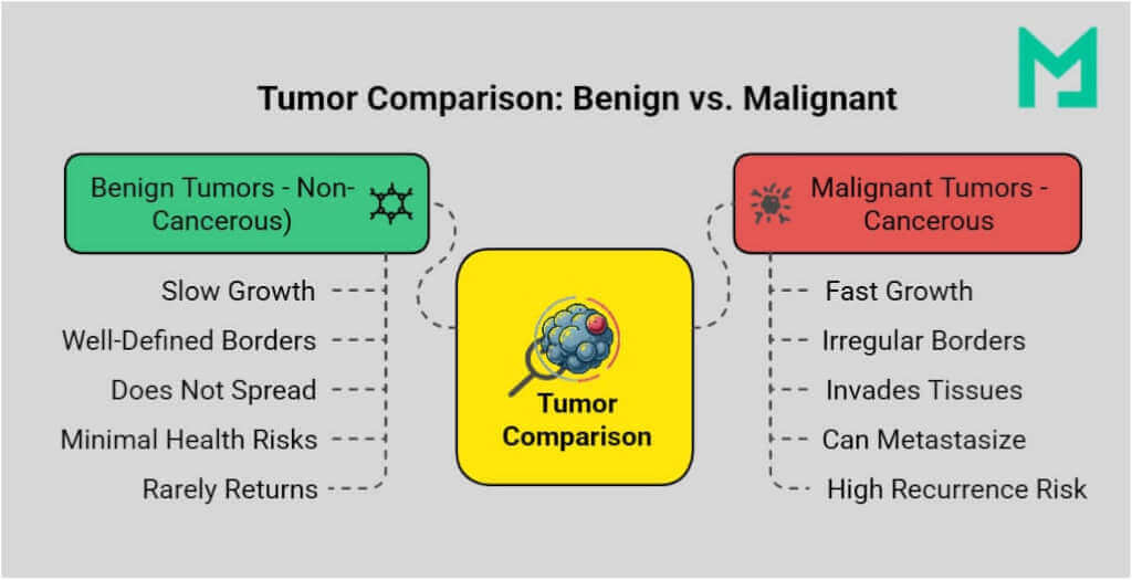
Benign tumors are non-cancerous, grow slowly, have well-defined borders, and do not spread, making them usually harmless. In contrast, malignant tumors are cancerous, grow rapidly, have irregular borders, invade nearby tissues, and can metastasize, necessitating urgent medical treatment.
Let’s discover benign vs malignant, how they’re diagnosed, and why early detection saves lives.

Benign vs Malignant: A Quick Peek
The most crucial distinction in tumors is whether they are benign (non-cancerous) or malignant (cancerous).
A benign tumor is non-cancerous, grows slowly, has well-defined borders, and does not invade or spread to other parts of the body, making it generally harmless. In contrast, a malignant tumor is cancerous, grows rapidly, has irregular borders, invades nearby tissues, and can spread (metastasize) to distant organs, requiring immediate medical treatment.
Benign vs malignant: 6 ways doctors describe the difference
Benign vs malignant tumors are typically separated by six practical differences:
- Recurrence: Benign tumors are less likely to recur after removal, whereas malignant tumors are more likely to recur.
- Growth: Benign tumors grow slowly; malignant tumors often grow faster.
- Borders: Benign tumors usually have smooth, well-defined borders, and malignant tumors often have irregular edges.
- Invasion: Benign tumors stay local, malignant tumors invade nearby tissue.
- Spread: Benign tumors do not metastasize; malignant tumors can spread through the blood or lymph nodes.
- Cell appearance: Benign cells look more like normal cells; malignant cells often look abnormal under a microscope.
Here’s a quick comparison to highlight their key differences:
| Feature | Benign | Malignant |
|---|
| Nature | Non-cancerous | Cancerous |
| Growth rate | Usually slow | Often faster |
| Borders | Well-defined | Irregular |
| Invasion | Does not invade nearby tissues | Invades nearby tissues |
| Metastasis | Does not spread | Can spread (metastasize) |
| Recurrence | Less likely after removal | More likely to return |
Let’s learn in detail how benign tumors are different from malignant tumors.
What Is The Difference Between Malignant And Benign?
A tumor is an abnormal mass of cells that grows uncontrollably when the body’s natural cell division goes awry. It can form in various body parts, such as the skin, organs, bones, and soft tissues. The impact of a tumor depends on factors such as its type, location, and growth patterns.
Tumors are classified into two main categories based on their ability to spread and their potential health risks:
- Benign Tumors
- Malignant Tumors
Benign Tumor
A benign tumor is a non-cancerous growth that forms when cells multiply abnormally but do not invade nearby tissues or spread to other parts of the body. Unlike malignant tumors, benign tumors grow slowly, remain localized, and have well-defined borders.
Benign tumors are generally harmless. However, if they grow large enough to press on organs, nerves, or blood vessels, they can cause complications.
While benign tumors are not life-threatening, they may sometimes require medical attention. Some benign tumors can resemble cancerous ones, making proper diagnosis crucial.
Characteristics of Benign Tumors
- Non-cancerous: Does not spread to other parts of the body.
- Slow-growing: Can remain unchanged for years.
- Well-defined borders: The tumor is encapsulated, keeping it separate from surrounding tissue.
- Does not invade nearby tissues: It stays confined to one location.
- Cells appear normal: Cells resemble healthy tissue rather than being irregular or mutated.
- Rarely recurs after removal: Benign tumors typically do not grow back once surgically removed.
Common Types of Benign Tumors
Benign tumors can develop in various tissues and organs of the body. Some common types include:
- Lipomas: Soft, fatty tissue tumors that develop under the skin, often on the arms, neck, or back.
- Adenomas: Tumors in glandular tissue, such as in the liver, colon, or thyroid. Examples include hepatic adenomas (in the liver) and colonic polyps (in the colon).
- Fibromas: Tumors that grow in connective or fibrous tissue, often seen in the uterus (uterine fibroids) or skin.
- Hemangiomas: Made up of abnormal blood vessels, commonly seen in infants, often appearing as a red birthmark.
- Nevi (Moles): Clusters of pigmented skin cells that form a mole, which can occasionally become cancerous over time.
When Should a Benign Tumor Be Removed?
While most benign tumors do not require treatment, some cases may necessitate surgical removal, especially when:
- Causes discomfort or pain: Presses on nearby tissues or organs.
- Obstructs vital functions: Can interfere with breathing or digestion.
- Risk of malignancy: Some benign tumors may become cancerous, prompting removal.
- Hormonal imbalances: Can disrupt hormone production and affect health.
- Affects appearance/self-esteem: Removal may be preferred for cosmetic reasons.

Malignant Tumor
A malignant tumor is a cancerous growth resulting from uncontrolled cell division, characterized by aggressive and invasive behavior. Unlike benign tumors, malignant tumors can invade surrounding tissues and metastasize to distant body parts via the bloodstream or lymphatic system.
Malignant tumors proliferate, often outpacing the body’s ability to regulate or remove them. This uncontrolled growth can disrupt organ function, destroy healthy cells, and lead to severe complications if not treated promptly.
Characteristics of Malignant Tumors
- Cancerous and aggressive: Grows quickly and spreads if left untreated.
- Invades surrounding tissues: Infiltrates nearby healthy tissues, making them difficult to remove completely.
- Can metastasize: Cancer cells can break away from the primary tumor and travel through the bloodstream or lymphatic system to form new tumors in other parts of the body.
- Cells appear abnormal: Under a microscope, malignant cells look irregular in size and shape.
- Recurrence risk: Even after treatment, cancer can return, mainly if not completely eradicated.
Common Types of Malignant Tumors
Malignant tumors can develop in various tissues and organs throughout the body. Some of the most common types include:
- Carcinomas: The most common type of cancer, originating in epithelial cells (the lining of organs and skin). Examples include:
- Lung cancer
- Breast cancer
- Colon cancer
- Skin cancer (melanoma)
- Sarcomas – Cancer arising from connective tissues, such as:
- Osteosarcoma (bone cancer)
- Liposarcoma (fat tissue cancer)
- Rhabdomyosarcoma (muscle cancer)
- Leukemias – Blood cancers that affect the bone marrow, leading to an overproduction of abnormal white blood cells. Examples include:
- Acute lymphoblastic leukemia (ALL)
- Chronic myeloid leukemia (CML)
- Lymphomas – Cancers affecting the lymphatic system are crucial for immune function. Examples include:
- Hodgkin’s lymphoma
- Non-Hodgkin’s lymphoma
Why Are Malignant Tumors Dangerous?
Malignant tumors pose serious health risks because of their ability to spread, destroy tissues, and resist treatment.
- Metastasis: Malignant tumors can spread to vital organs such as the brain, liver, or lungs, complicating treatment.
- Tissue Damage: They harm healthy cells, impair organ function, and may lead to severe pain and life-threatening complications.
- Recurrence: Cancer can return after treatment, and some may become resistant, necessitating ongoing management.
Benign vs. Malignant Ulcer
While tumors are often the focus of benign and malignant cancer screenings, other abnormal growths like ulcers may also require medical evaluation.
Benign ulcers, such as peptic ulcers, result from infections or inflammation and heal with treatment. However, malignant ulcers, like gastric cancer-related ulcers, persist, grow irregularly, and may bleed.
Advanced imaging and biopsy analysis help distinguish benign and malignant ulcers, ensuring timely treatment.
How Are Benign and Malignant Tumors Diagnosed?
Proper diagnosis is crucial to distinguish between benign and malignant tumors. Doctors utilize AI-backed imaging tests, biopsies, and blood tests to evaluate the tumor and decide on the best treatment.
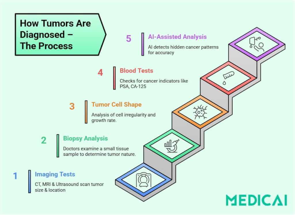
Medical tests for tumor diagnosis include:
Imaging Tests
Visualize the tumor’s size, shape, and location, and check for spreading.
- X-rays: Detect tumors in bones and soft tissues.
- Ultrasound: Examines tumors in organs like the liver and thyroid.
- CT Scan: Creates detailed 3D images of tumors and surrounding areas.
- MRI: Provides high-resolution images of soft tissue tumors in the brain, muscles, or joints.
- PET Scan: Detects cancerous activity by analyzing cell metabolism.
- Ultrasonographic Scoring System: This novel approach combines vascularity index (VI), maximal shear velocity (MSV), and tumor size to distinguish benign from malignant tumors with 93.6% sensitivity and 79.2% specificity. This non-invasive tool significantly enhances diagnostic precision and reduces unnecessary biopsies.
Advancements in medical imaging have also improved how teams review and share tumor studies. When a case needs follow-up, second opinions, or tumor board input, a cloud PACS and DICOM viewer can make it easier to pull up prior scans, compare studies side-by-side, and securely share the same imaging context. Medicai is one example of a platform used for that kind of imaging workflow.
How imaging teams use PACS to support benign vs malignant workups
Imaging is often the first clue that a tumor appears benign or suspicious, but the diagnostic workup only moves quickly when the images, priors, and report are easy to access and share.
PACS (Picture Archiving and Communication System) is the system radiology teams use to store, view, compare, and distribute imaging studies. In a benign vs malignant workup, PACS supports six practical steps:
- Compare priors without delay: Radiologists often need the most recent CT, MRI, ultrasound, or PET study to determine whether a mass is stable, growing, or changing in a concerning way.
- Keep multi-modality context in one place: Suspicious findings often prompt multiple modalities. PACS helps teams review the “full picture” instead of treating each scan as a separate event.
- Standardize how findings are described: Structured reporting helps capture lesion size, location, growth patterns, and recommended follow-up in a consistent format, so the next clinician does not miss key details.
- Support second opinions and tumor board review: Complex cases move faster when the same study can be reviewed by multiple clinicians without file transfers or missing context.
- Protect access while enabling care coordination: Imaging access has to be controlled and auditable, especially when multiple departments or external specialists are involved.
- Reduce rework and repeat imaging: When priors and reports are hard to retrieve, teams end up repeating scans. Clean access and sharing reduce unnecessary repetition.
Cloud PACS and web-based DICOM viewers are designed for these workflows. Medicai is an example of a platform that supports cross-site viewing and secure collaboration when multiple clinicians need the same imaging context.
Biopsy (Tissue Sample Analysis)
Doctors examine a small tissue sample under a microscope to distinguish between benign and malignant tumors for unusual growth and cancer cells.
Biopsies involve different methods to collect tissue, including needle biopsies with a thin needle, surgical biopsies that remove tumor portions, and endoscopic biopsies using a camera for internal sampling.
Blood Tests for Tumor Markers
Certain cancers release biomarkers that assist in diagnosis. PSA indicates prostate cancer, CA-125 suggests ovarian cancer, AFP can mark liver cancer, and CEA may indicate colon or lung cancer.
How to Tell if a Tumor is Cancerous?
The only way to confirm malignancy is through microscopic examination of tumor cells. Doctors look for the following in cancerous tumors:
- Irregularly shaped cells: Cancer cells appear abnormal and disorganized compared to normal, uniform cells.
- Rapid cell division: Malignant tumors have a high growth rate, seen under a microscope.
- High vascularity: Malignant tumors show a vascularity index (VI) >10%, significantly higher than benign tumors (VI ~2.4%).
- Abnormal tissue stiffness: Malignant tumors exhibit higher shear velocity (≥8.3 m/sec) compared to benign tumors (6.1 m/sec).
- Invasion of surrounding tissue: If cancer cells are spreading into adjacent structures, it indicates malignancy.
- Genetic mutations: Advanced tests, such as molecular profiling, can identify cancer-related mutations.
Some teams use AI tools to highlight suspicious patterns on imaging and keep those outputs attached to the study for review, but clinician judgment and, when needed, biopsy-based pathology still determine whether a tumor is malignant.

Treatment Options: Benign vs. Malignant Tumors
Treatment for tumors varies significantly based on whether they are benign (non-cancerous) or malignant (cancerous).
Benign Tumor Treatment
Most benign tumors do not require treatment, but intervention may be necessary if they cause discomfort or pain, affect normal function, or have the potential to turn cancerous.
Treatment options for benign tumors include:
- Monitoring: Small, symptom-free tumors may require regular check-ups.
- Surgical Removal: Growing or problematic tumors can be surgically excised.
- Medications: Benign tumors, like hormone-secreting adenomas, may be treated with medication.
Malignant Tumor Treatment
Malignant tumors require urgent medical intervention to prevent the spread of cancer. Treatment varies depending on the cancer type, stage, and the patient’s health condition.
Common treatments for malignant tumors include:
- Surgery: Primary treatment for solid tumors, aimed at complete removal of cancer.
- Chemotherapy: Uses drugs to kill or halt cancer cell growth, often for blood cancers or metastatic disease.
- Radiation Therapy: Employs high-energy rays to shrink tumors and destroy cancer cells.
- Immunotherapy: Enhances the immune system to combat cancer.
- Targeted Therapy: Targets specific genetic mutations in cancer cells, sparing healthy ones. Second-generation KRAS inhibitors are expanding targeted therapy options for lung, colorectal, and pancreatic cancers.
- Hormone Therapy: For hormone-sensitive cancers like breast and prostate cancer.
Can a Benign Tumor Become Malignant?
Benign tumors are usually non-cancerous and pose little health risk. However, some can undergo malignant transformation over time due to factors like genetic mutations, chronic inflammation, and prolonged exposure to carcinogens.
Examples of benign tumors that may become malignant include—
- Colonic Polyps to Colorectal Cancer: Adenomatous polyps can develop into colon cancer if untreated.
- Moles (Nevi) to Melanoma: Most moles are harmless, but some atypical moles (dysplastic nevi) may evolve into skin cancer (melanoma) over time.
- Leiomyomas (Fibroids) to Leiomyosarcomas: Benign uterine fibroids may develop into a malignant tumor known as leiomyosarcoma.
- Papillomas to Certain Cancers: Some benign papillomas, particularly those caused by HPV (Human Papillomavirus), can increase the risk of cervical, throat, or bladder cancer.
- Benign vs Malignant Ovarian Cyst: Benign ovarian cysts often resolve on their own, while malignant ones require immediate medical attention.
Next steps: what to do if a scan shows a tumor
A scan can show that a tumor exists, but a scan alone cannot confirm cancer. The next steps are to confirm the tumor type and its aggressiveness and determine the plan that makes sense for your case.
What to do next, as a patient or caregiver
- Ask for the radiology report and the image access plan: Get a copy of the report and ask how you or your doctor will access the images for follow-up and second opinions.
- Ask what the finding means in plain language: Ask whether the tumor looks more consistent with a benign pattern or a suspicious pattern, and what features led to that impression.
- Confirm the recommended next test and timeline: Typical next steps include follow-up imaging, a referral to a specialist, or a biopsy. Ask what is recommended and when it should happen.
- Ask what changes the plan: Ask what symptoms or changes would require faster evaluation.
What imaging teams should keep tight, so care does not stall
- Ensure priors are available for side-by-side review: The fastest reads happen when relevant prior exams are already accessible.
- Make sharing and review simple for specialists: Second opinions and tumor boards require the same study context, not screenshots or incomplete exports.
- Keep reporting consistent and easy to retrieve: Clear, standardized findings and follow-up recommendations reduce missed handoffs.
This is where an imaging platform matters. A cloud PACS workflow, such as Medicai, can help teams keep studies accessible for collaboration and follow-up without relying on discs or manual transfers.


Related Articles


Lets get in touch!
Learn more about how Medicai can help you strengthen your practice and improve your patients’ experience. Ready to start your Journey?
Book A Free Demo