Mammography PACS & Tomosynthesis: Solving the "Heavy Data" Problem
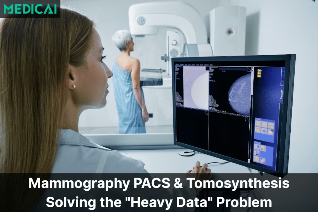
In the last decade, breast imaging has undergone a massive technological leap. We moved from 2D Digital Mammography (FFDM) to Digital Breast Tomosynthesis (DBT).
Clinically, this is a victory—detection rates are up, and recalls are down. IT-wise, it is a crisis.
A standard 4-view 2D mammogram is roughly 50MB. A comprehensive 3D Tomosynthesis study with C-View and priors can easily exceed 600MB to 1GB per patient.
For many Radiology IT Directors, this data explosion has broken their existing infrastructure. Storage costs are skyrocketing, and radiologists are left staring at loading bars because their standard “Cloud Viewer” cannot stream 1GB files fast enough over a standard internet connection.
This guide explores the specific technical requirements for a modern Mammography PACS and why a Hybrid VNA approach is the only sustainable path forward.
The Unique Requirements of a 3D Mammo Viewer
You cannot treat a mammogram like a chest X-ray. The workflow is highly specialized, and a generic PACS viewer will simply fail to meet the radiologist’s needs. To support high-volume breast imaging, your viewer needs three non-negotiable attributes:

1. Advanced Hanging Protocols
In breast imaging, comparison is everything. A radiologist doesn’t just “open” an image; they need the system to automatically arrange the current CC/MLO views next to the prior year’s CC/MLO views.
- The DBT Challenge: With Tomosynthesis, the protocol must also manage the “Cine Loop” stack. It needs to auto-sync the scrolling so that as the radiologist scrolls through the Left MLO 3D volume, the Right MLO scrolls in unison.
2. Instant Cine Mode (No Buffering)
Detecting microcalcifications requires smooth, high-frame-rate navigation through the 3D slice stack.
- The Latency Trap: If your cloud PACS tries to download the entire 600MB file before playing, the doctor waits 45 seconds per patient. If it “buffers” during scrolling, diagnostic confidence drops. The viewer must use Server-Side Rendering or Adaptive Streaming to play instantly.
3. Vendor-Neutral Display
Historically, Hologic or GE dimensions were viewed on dedicated, proprietary workstations. A modern Mammography PACS must be vendor-agnostic, allowing you to view images from a Hologic Selenia, GE Senographe, or Siemens Mammomat on a single, unified reading station.
The Storage Crisis: Why “Pure Cloud” Struggles
If you perform 40 tomosynthesis studies a day, you are generating roughly 20GB of new data daily.
- On-Premise Issue: You will fill up your local SAN (Storage Area Network) in 12 months, forcing an expensive hardware upgrade.
- Cloud Issue: As discussed in our Hybrid PACS Technical Guide, pushing 20GB/day over a standard ISP connection can choke your network, and pulling it back down for reading creates latency.
The Solution: Medicai’s Hybrid VNA for Breast Imaging
Medicai solves the “Heavy Data” problem by decoupling storage from viewing using our Hybrid Edge Architecture.
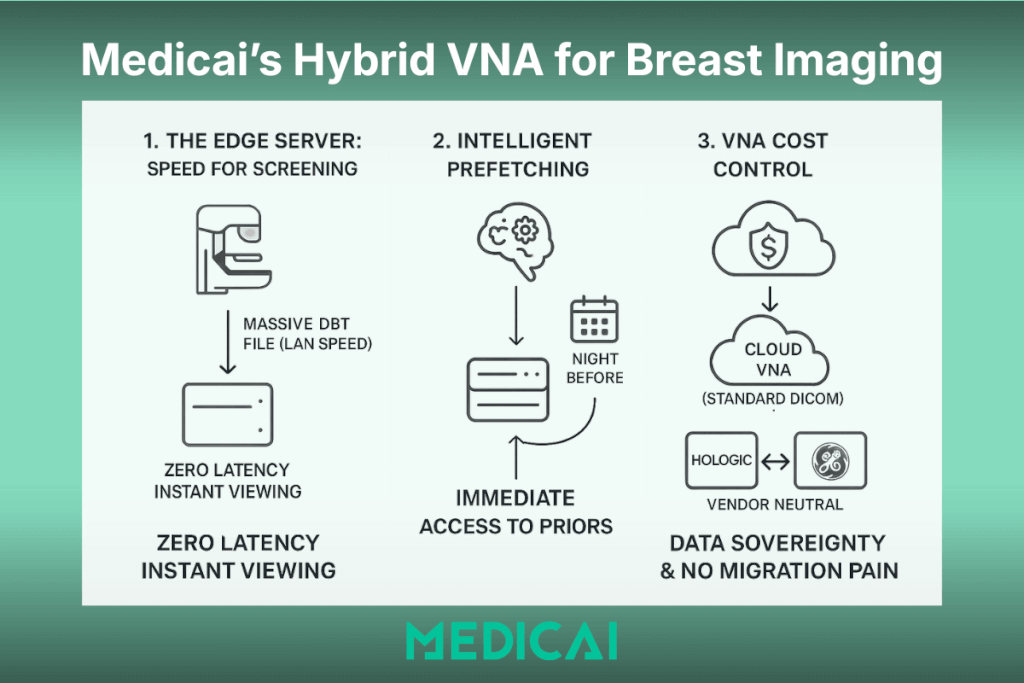
1. The Edge Server: Speed for Screening
We place a small Edge Server locally at your breast imaging center.
- The Workflow: When a patient is scanned, the massive DBT file goes directly to the local Edge Server (over your fast LAN).
- The Benefit: Your radiologists experience Zero Latency. The large 3D files open instantly because they are actually pulled from the local server, even though they use a web-based viewer.
2. Intelligent Prefetching
What about priors? If a patient returns for their annual screening, Medicai’s AI automatically pre-fetches their prior 3D exams from the cloud to the local Edge Server the night before their appointment.
- Result: The radiologist has immediate access to historical comparisons without waiting for a cloud download.
3. VNA Cost Control
Instead of buying expensive proprietary storage from your modality vendor, Medicai stores your massive DBT archives in a cost-effective Cloud VNA.
- Data Sovereignty: We store images in standard DICOM format. If you ever switch hardware vendors (e.g., from Hologic to GE), you don’t need a painful Data Migration project. Your archive is neutral and ready for the new modality.
Conclusion
Breast imaging saves lives, but it also creates the biggest data footprint in radiology. Do not let “File Size” dictate your efficiency.
By moving to a Hybrid Mammography PACS, you get the best of both worlds: the instant speed of on-premise reading for your doctors, and the infinite, low-cost scalability of the cloud for your IT budget.
Is your network ready for 3D Tomo? Don’t let buffering slow down your screening program.


Related Articles

Lets get in touch!
Learn more about how Medicai can help you strengthen your practice and improve your patients’ experience. Ready to start your Journey?
Book A Free Demo