Contrast MRI: From Prep to Image Clarity

What if your MRI scan could reveal hidden tumors or tiny vessel leaks in stunning detail?
Contrast MRI acts like a spotlight, bringing unseen abnormalities into clear view.
MRI with contrast uses intravenous gadolinium chelates to shorten T1 relaxation, making lesions, vessels, and inflammation appear brighter on T1-weighted images. This enhanced visibility boosts sensitivity, detects tiny abnormalities, and enables precise treatment planning.
Dive into our in-depth guide to discover preparation tips, safety measures, and clinical indications in contrast MRI.

What Is MRI With Contrast?
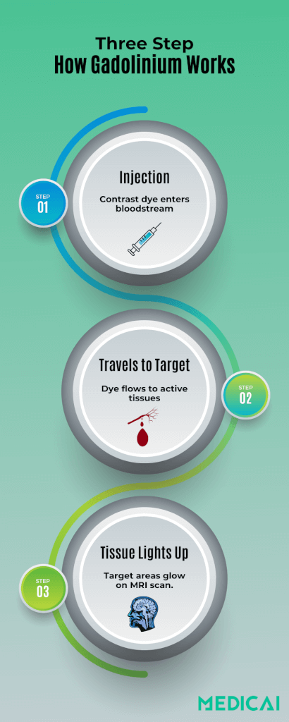
An MRI with contrast is a specialized scan where a harmless dye, typically a gadolinium-based contrast agent (GBCA), is injected into the bloodstream before the images are taken. This dye makes certain tissues, such as tiny tumors, inflamed areas, or leaky blood vessels, stand out more clearly in the pictures.
By “lighting up” these spots, contrast-enhanced MRI helps doctors see details that might be missed on a standard scan.
What’s a GBCA?
A Gadolinium-Based Contrast Agent (GBCA) is the most common dye used for contrast MRI. It combines gadolinium—a metal that strongly affects MRI signals—with molecules called chelates that keep it safe for your body.
Once injected, the GBCA travels to areas with extra blood flow or damaged vessel walls. There, it shortens the T1 relaxation time of nearby hydrogen atoms, so those regions appear brighter on T1-weighted images. This brightening makes small abnormalities far more visible.
GBCAs light up images by
- Speeding Relaxation: After the MRI’s radio pulse, hydrogen atoms emit signals. Gadolinium makes them relax and send signals faster, boosting their brightness.
- Selective Retention: Normal tissue washes out the agent quickly, while lesions or inflammation often hold it longer. The difference in brightness helps radiologists pinpoint problems.
Linear vs. Macrocyclic Agents
Contrast chelates come in two shapes:
- Linear GBCAs use an open-chain molecule wrapped around gadolinium. They enhance images well but can release tiny amounts of free gadolinium over time.
- Macrocyclic GBCAs form a tight, ring-shaped “cage” around gadolinium. This secure binding significantly reduces the chance of gadolinium escaping into tissues.
Because macrocyclic agents hold gadolinium more firmly, they carry a lower risk of long-term metal accumulation in the brain and rare side effects, such as nephrogenic systemic fibrosis. For this reason, doctors often choose macrocyclic GBCAs for children, pregnant patients, and anyone with kidney concerns.
Clinical Indications of Contrast MRI
Contrast-enhanced MRI adds vital detail by “lighting up” abnormal areas that ordinary scans might miss.
Tumor Evaluation
When a tumor grows new blood vessels, it absorbs contrast dye more readily than healthy tissue. In the brain, this makes gliomas—aggressive tumors—appear bright against normal gray and white matter.
In the liver, small metastases appear as glowing spots, enabling surgeons and oncologists to create precise treatment plans.
Infection & Inflammation
Infected or inflamed tissue often becomes leaky and flooded with blood, trapping the contrast agent. For example, in osteomyelitis (a bone infection), the outline of an abscess is visible, guiding surgical drainage.
In Crohn’s disease, contrast highlights active loops of inflamed intestine, pinpointing areas that may need medicine or surgery.
Vascular Mapping
Blood vessels themselves can be the target of an MRI. In contrast, arteries and veins fill with dye, enabling three-dimensional views of aneurysms or arteriovenous malformations (AVMs). These detailed maps help interventional radiologists plan coil placements or surgical repairs without guesswork.
Demyelinating Disease
In conditions like multiple sclerosis, active plaques disrupt the blood–brain barrier and absorb contrast. On T1-weighted images, these bright lesions reveal sites of new or ongoing damage. Tracking their number and size over time helps neurologists adjust therapies to slow disease progression.

Chemistry & Safety of Contrast Agents
Two major safety concerns are the potential for gadolinium to remain in tissues and the regulation of different agents by health authorities.
Gadolinium Deposition in the Brain
Research shows that small amounts of gadolinium deposition occur in T1-hyperintensity areas after repeated scans. Linear agents tend to leave more of these trace deposits than macrocyclic ones.
To date, no clear harm has been linked to these tiny residues. Still, many doctors choose macrocyclic dyes for patients who require multiple exams—especially children and those with kidney issues—to minimize potential deposits as much as possible.
Global Regulatory Guidance
The first gadolinium-based contrast agents (GBCAs) were approved by the FDA in 1998. However, because of deposition concerns, different regions have taken varied approaches:
European Union: Has restricted or withdrawn most linear GBCAs, favoring macrocyclic agents across the board.
United States (FDA): Continues to approve both linear and macrocyclic GBCAs but recommends that high-risk groups—patients with poor kidney function, children, and those needing frequent scans—receive macrocyclic agents whenever possible.
The Patient Experience: Step-by-Step
Before your MRI scan begins, you’ll go through a few simple steps to make sure everything goes smoothly and safely.
1. Scheduling & Screening
When you book your MRI with contrast, the clinic will ask about your kidney health, allergies, implants, and pregnancy. You may need a recent blood test (eGFR) to confirm your kidneys can clear the dye safely.
2. Preparation
For abdominal scans, you’ll fast for about 4 hours beforehand. Otherwise, you can eat normally but stay hydrated. Wear loose, metal-free clothing and leave jewelry at home. Let the staff know if you feel nervous—many centers offer mild sedation.
3. IV Placement & Scout Images
A nurse or technologist will insert a small IV cannula in your arm. They’ll run a few quick “scout” images without contrast to plan the detailed scan sequences.
4. Contrast Injection & Scanning
The gadolinium agent is injected through your IV over 5–20 seconds. You might feel a cool flush or metallic taste for a moment. The scanner then captures images in different phases (arterial, venous, delayed), usually adding around 10 minutes to the exam.
Throughout, you’ll lie still and hear gentle tapping sounds.
5. Aftercare & Follow-Up
Once the scan ends, the IV is removed, and you can go home. Drinking an extra 1–2 liters of water over the next 24 hours helps flush the agent from your body.
You can resume normal activities right away unless you’ve had sedation.

Benefits of MRI with Contrast
Contrast-enhanced MRI uncovers details that a standard scan can miss.
Increased Sensitivity and Early Detection
Contrast-enhanced MRI can reveal lesions as small as 2–3 mm that non-contrast scans often miss. This heightened sensitivity helps doctors to spot early-stage tumors or subtle inflammation, allowing treatment to begin when it’s most effective.
Platforms like Medicai offer automated image segmentation that highlights enhanced regions in seconds, so radiologists spot tiny lesions faster.
Precise Treatment Planning
By clearly defining tumor margins, contrast MRI shows exactly where abnormal tissue ends and healthy tissue begins. Surgeons use these images to plan more accurate resections, and oncologists monitor contrast uptake patterns to adjust chemotherapy or radiation with confidence.
Consolidated Imaging and Cost Savings
A single contrast MRI exam can answer multiple clinical questions—reducing the need for separate non-contrast and follow-up scans. Fewer repeat studies streamline patient care, cut the overall costs of MRI, and shorten the time to a definitive diagnosis.
Improved Patient Experience
Less guesswork means fewer appointments and less waiting. Patients benefit from a one-stop exam that provides comprehensive information, leading to quicker treatment decisions and reduced anxiety.
By combining advanced algorithms with intuitive dashboards, Medicai enhances diagnostic confidence, reduces repeat scans, and enables your team to focus on delivering personalized patient care.
Risks, Side Effects & Contraindications
MRI with contrast is safe for most people, but like any medical procedure, it carries some risks.
| Severity | Typical Onset | Frequency | Examples |
| Mild | Minutes | 1–5% | Nausea in children, metallic taste, local warmth |
| Moderate | Minutes–hours | 0.1–0.5% | Urticaria, bronchospasm |
| Severe (anaphylaxis) | Immediate | 0.01–0.03% | Hypotension, airway edema |
| NSF | Days–months | <0.01% overall; up to 4% in GFR <30 mL/min with certain linear agents | Skin thickening, joint contractures |
| Brain deposition | Months–years | Detectable, but clinical impact unknown | Dentate nucleus hyperintensity |
Special Populations
- Renal impairment – Use macrocyclic GBCA at standard dose or avoid contrast if GFR <30 mL/min; dialysis within 2 hours removes ≈70% of gadolinium.
- Pediatrics – Acute reaction rates are lower than in adults, but macrocyclic agents are preferred to minimize brain deposition risk.
- Pregnancy – GBCAs cross the placenta; use only if essential, opting for macrocyclic agents and the lowest effective dose.
- Breastfeeding – Less than 0.04% of the dose appears in milk; continuation is considered safe, but 24-hour pump-and-discard is offered for anxious mothers.
MRI With Or Without Contrast: What are The Differences
Contrast MRI uses gadolinium to “light up” tiny lesions and vessels, thereby enhancing detection. On the other hand, non-contrast MRI is quicker, cheaper, and ideal for routine imaging.
| Feature | Without Contrast | With Contrast |
| Lesion Detection | Good for large masses | Detects lesions as small as 2–3 mm |
| Vascular Detail | Flow-sensitive sequences only | Bright-blood imaging for clear 3D vessel maps |
| Inflammation vs. Scar | T2 hyperintensity only | Highlights active inflammation and capillary leak |
| Scan Time | 20–40 minutes | 25–50 minutes (adds 5–10 min for contrast phases) |
| Cost | Lower | Higher (contrast agent adds ≈ US$75–200) |
| Repeat Scans Needed | May require follow-up non-contrast studies | Often, one study answers multiple questions |
| Patient Preparation | No special prep beyond metal screening | May need fasting and renal function tests beforehand |
Conclusion
MRI with contrast brings hidden details into focus, enabling early detection, precise treatment planning, and streamlined patient care. By selecting the safest gadolinium agents and adhering to best-practice screening protocols, clinicians maximize diagnostic value while minimizing risk.
With Medicai’s intelligent segmentation, quantitative perfusion analysis, and secure PACS integration, your workflow becomes faster and more reliable. Now, you can deliver confident, personalized diagnoses with efficiency and ease.


Related Articles


Lets get in touch!
Learn more about how Medicai can help you strengthen your practice and improve your patients’ experience. Ready to start your Journey?
Book A Free Demo